 Back
BackChapter 19: The Kidneys – Structure, Function, and Physiology
Study Guide - Smart Notes
 Tailored notes based on your materials, expanded with key definitions, examples, and context.
The Kidneys: Structure, Function, and Physiology
Overview
The kidneys are essential organs in the urinary system, responsible for maintaining homeostasis by regulating fluid balance, electrolyte concentrations, and waste excretion. This chapter provides an integrated understanding of kidney anatomy, nephron function, and the physiological processes of filtration, reabsorption, secretion, and excretion.
Functions of the Kidneys
Main Physiological Roles
Regulation of Extracellular Fluid Volume and Blood Pressure: The kidneys adjust urine output to maintain blood volume and pressure.
Regulation of Osmolarity: By controlling water and solute excretion, kidneys keep plasma osmolarity near 290 mOsM.
Maintenance of Ion Balance: The kidneys regulate key ions such as Na+, K+, and Ca2+.
Homeostatic Regulation of pH: Kidneys excrete H+ and reabsorb HCO3- to maintain acid-base balance.
Excretion of Wastes: Metabolic wastes (urea, creatinine), toxins, and drugs are eliminated in urine.
Production of Hormones: Kidneys secrete erythropoietin (stimulates RBC production), renin (regulates blood pressure), and activate vitamin D.
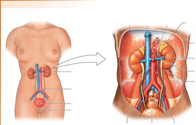
Anatomy of the Urinary System
Major Structures
Kidneys: Paired, retroperitoneal organs that filter blood and produce urine.
Ureters: Muscular tubes transporting urine from kidneys to the bladder.
Urinary Bladder: Stores urine until micturition (urination).
Urethra: Conducts urine from the bladder to the external environment; differs in length and position between males and females.
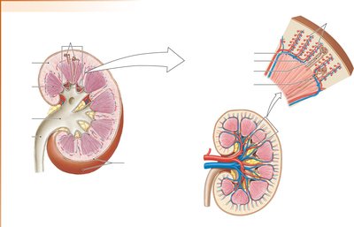
Kidney Structure
Cortex: Outer region containing 80% of nephrons (cortical nephrons).
Medulla: Inner region with 20% of nephrons (juxtamedullary nephrons).
Renal Pelvis: Collects urine from nephrons before passing it to the ureter.
Vascular Elements: Blood enters via the renal artery, passes through afferent arterioles, glomerulus, efferent arterioles, and peritubular capillaries/vasa recta (portal system).
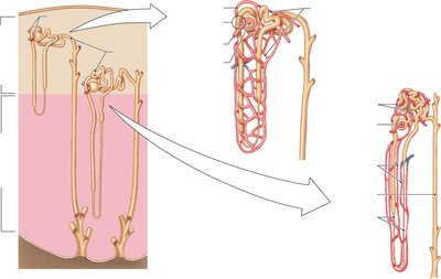
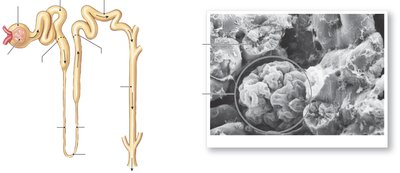
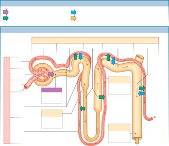
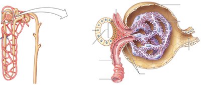
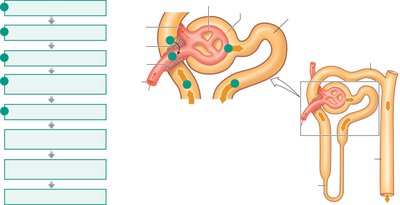
Nephron Anatomy
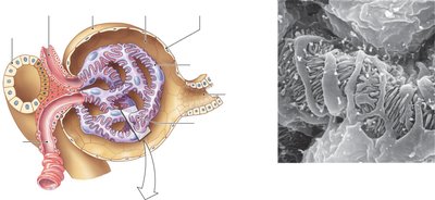
Renal Corpuscle: Composed of Bowman’s capsule and glomerulus; site of filtration.
Proximal Tubule: Major site of reabsorption.
Loop of Henle: Descending and ascending limbs; establishes osmotic gradient.
Distal Tubule: Further reabsorption and secretion.
Collecting Ducts: Final site for urine concentration and water reabsorption.
Juxtaglomerular Apparatus: Specialized region where the ascending limb contacts afferent and efferent arterioles; important for regulating GFR and blood pressure.
Overview of Kidney Function
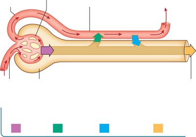
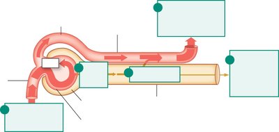
Basic Renal Processes
Filtration: Movement of fluid from blood into the nephron lumen at the renal corpuscle. The filtered fluid is called filtrate.
Reabsorption: Return of useful substances from the filtrate back into the blood, mainly in the proximal tubule and peritubular capillaries.
Secretion: Active transport of additional substances from blood into the nephron tubule.
Excretion: Removal of final urine from the body.
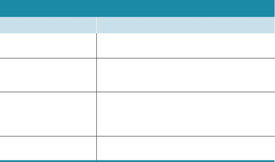
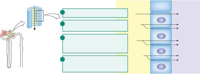
Summary Table: Nephron Segments and Functions
Segment of Nephron | Main Functions |
|---|---|
Renal corpuscle (glomerulus + Bowman’s capsule) | Filtration of mostly protein-free plasma from capillaries into the capsule |
Proximal tubule | Isosmotic reabsorption of organic nutrients, ions, and water; secretion of metabolites and xenobiotics |
Loop of Henle | Reabsorption of ions in excess of water; creates dilute fluid in the lumen; contributes to medullary osmotic gradient |
Distal nephron (distal tubule + collecting duct) | Regulated reabsorption of ions and water; salt and water balance; pH homeostasis |
Filtration
Filtration Fraction and Barriers
Filtration Fraction: Percentage of renal plasma flow that becomes filtrate (about 20%).
Filtration Barriers: Consist of glomerular capillary endothelium (fenestrated), basement membrane, and podocyte filtration slits.
Forces Driving Filtration
Capillary Blood Pressure (PH): Hydrostatic pressure (~55 mm Hg) favors filtration.
Capillary Colloid Osmotic Pressure (π): Due to plasma proteins (~30 mm Hg), opposes filtration.
Capsule Fluid Pressure (Pfluid): Hydrostatic pressure in Bowman’s capsule (~15 mm Hg), opposes filtration.
Net Filtration Pressure:
Glomerular Filtration Rate (GFR)
Definition: Volume of fluid filtered per unit time (about 125 mL/min).
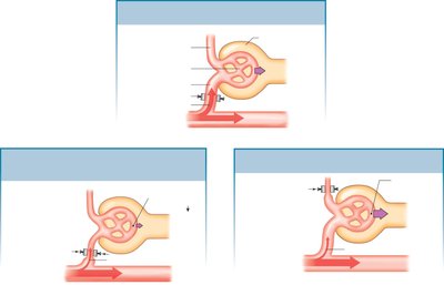
Regulation: Controlled by blood flow through renal arterioles. Increased afferent resistance decreases GFR; increased efferent resistance increases GFR.
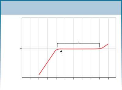
Autoregulation: Maintains constant GFR over a wide range of blood pressures (80–180 mm Hg).
Mechanisms of GFR Regulation
Myogenic Response: Vascular smooth muscle responds to pressure changes to maintain GFR.
Tubuloglomerular Feedback: Macula densa cells sense NaCl in filtrate and signal afferent arteriole to constrict or dilate.
Hormonal and Neural Control: Hormones and autonomic neurons adjust arteriole resistance.
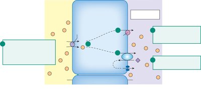
Reabsorption
Mechanisms and Pathways
Active and Passive Reabsorption: Most occurs in the proximal convoluted tubule (PCT).
Transcellular (epithelial) Transport: Solutes move through cells via membrane transporters.
Paracellular Pathway: Solutes move between cells through tight junctions.
Water Reabsorption: Follows solute reabsorption by osmosis.
Sodium-Linked Reabsorption
Na+ Reabsorption: Actively transported out of tubule cells by Na+-K+-ATPase.
Secondary Active Transport: Na+ gradient drives cotransport of glucose, amino acids, and other solutes via SGLT proteins.
Glucose Reabsorption: Glucose enters cells with Na+ and exits via GLUT transporters.
Renal Transport Maximum and Threshold
Saturation: Maximum rate of transport when all carriers are occupied.
Renal Threshold: Plasma concentration at which a substance first appears in urine (e.g., glucose in diabetes).
Secretion
Active Secretion
Definition: Active movement of molecules from extracellular fluid into nephron lumen.
Roles: Homeostatic regulation of K+ and H+; elimination of drugs, metabolites, and toxins.
Excretion
Final Urine Formation
Equation:
Clearance: Volume of plasma cleared of a substance per minute; used to estimate GFR.
Clinical Markers: Inulin (ideal but impractical) and creatinine (commonly used) are used to measure GFR.
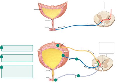
Micturition
Urine Storage and Elimination
Storage: Urine is stored in the bladder until micturition.
Control: Two sphincters regulate flow: internal (smooth muscle, involuntary) and external (skeletal muscle, voluntary).
Reflex: Stretch receptors in the bladder wall trigger a spinal reflex, causing bladder contraction and sphincter relaxation. The brainstem and cortex can override this reflex.
Additional info: This summary integrates textbook content and academic context to provide a comprehensive, exam-focused overview of kidney physiology, suitable for ANP college students.
