 Back
Chapter 2: The Cell – Structure, Function, and Life Cycle
Study Guide - Smart Notes
 Tailored notes based on your materials, expanded with key definitions, examples, and context.
Introduction to Cells
Overview of Cellular Biology
Cells are the fundamental structural and functional units of all living organisms. In the human body, trillions of cells perform specialized roles, forming tissues, organs, and systems. All cells arise from the division of preexisting cells and are responsible for carrying out all vital functions necessary for life.
Sex cells (germ cells): Sperm in males and oocytes in females, involved in reproduction.
Somatic cells: All other body cells that are not sex cells.
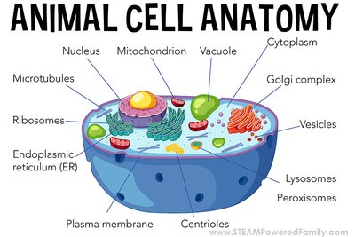
Cellular Anatomy
Major Components of the Cell
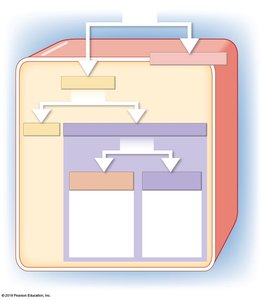
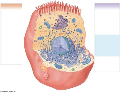
Each cell consists of three main components: the plasma membrane, cytoplasm (including cytosol and organelles), and the nucleus. Organelles are further classified as membranous or nonmembranous based on the presence of a surrounding membrane.
Plasma membrane: The outer boundary of the cell, regulating interactions with the environment.
Cytoplasm: The material within the cell, excluding the nucleus, containing cytosol and organelles.
Nucleus: The control center containing genetic material (DNA).
The Plasma Membrane
Structure and Function
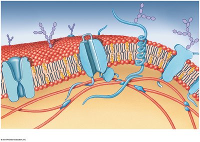
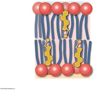
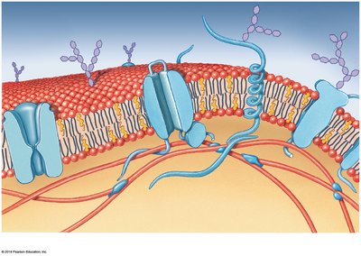
The plasma membrane is a selectively permeable barrier composed of a phospholipid bilayer with embedded proteins, glycolipids, and sterols. It provides structural support, mediates communication, and regulates the movement of substances into and out of the cell.
Physical barrier: Separates the internal environment from the external environment.
Regulation: Controls the exchange of materials.
Sensitivity: Responds to changes in the extracellular fluid.
Cell-to-cell communication: Facilitates adhesion and signaling.
Components of the Plasma Membrane
Phospholipids: Form the bilayer with hydrophilic heads facing outward and hydrophobic tails inward.
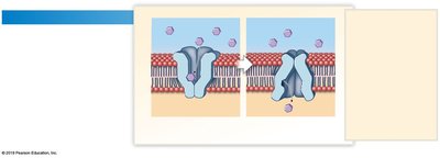
Proteins: Peripheral (surface) and integral (embedded); some form channels or gated channels for transport.
Glycolipids and glycoproteins: Present on the outer layer, forming the glycocalyx for cell recognition and signaling.
Sterols (e.g., cholesterol): Stabilize membrane structure.
Membrane Permeability: Passive and Active Processes
The plasma membrane's permeability determines how substances move across it. It can be impermeable, freely permeable, or selectively permeable. Transport mechanisms are classified as passive (no energy required) or active (energy required).
Passive processes: Diffusion, osmosis, facilitated diffusion
Active processes: Active transport, endocytosis (pinocytosis, phagocytosis, receptor-mediated), exocytosis
Passive Processes
Diffusion: Movement of molecules from high to low concentration (), down a concentration gradient.
Osmosis: Diffusion of water across a selectively permeable membrane toward higher solute concentration.
Facilitated diffusion: Transport of solutes via carrier proteins, following the concentration gradient.
Active Processes
Active transport: Movement of substances against their concentration gradient using carrier proteins and ATP (e.g., sodium-potassium pump).
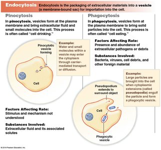
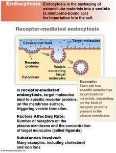
Endocytosis: Uptake of materials via vesicle formation. Includes pinocytosis (cell drinking), phagocytosis (cell eating), and receptor-mediated endocytosis.
Exocytosis: Release of intracellular materials by vesicle fusion with the plasma membrane.
Extensions of the Plasma Membrane
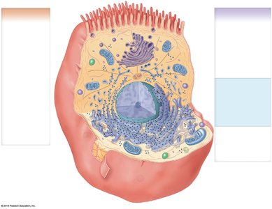
Microvilli: Fingerlike projections that increase surface area for absorption and aid in nutrient uptake.
The Cytoplasm
Cytosol and Organelles
The cytoplasm includes the cytosol (intracellular fluid) and organelles. The cytosol contains ions, proteins, nutrients, and inclusions. Organelles perform specialized cellular functions and are classified as nonmembranous or membranous.
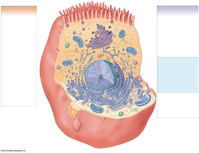
Nonmembranous organelles: Cytoskeleton, centrioles, cilia, flagella, ribosomes
Membranous organelles: Mitochondria, nucleus, endoplasmic reticulum, Golgi apparatus, lysosomes, peroxisomes
Nonmembranous Organelles
Cytoskeleton: Provides structural support and facilitates movement. Includes microfilaments (actin), intermediate filaments, thick filaments (myosin), and microtubules (tubulin).
Centrioles: Organize microtubules during cell division.
Cilia and Flagella: Surface projections for movement of fluids (cilia) or cell propulsion (flagella).
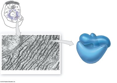
Ribosomes: Sites of protein synthesis; can be free in cytosol or attached to the endoplasmic reticulum.
Membranous Organelles
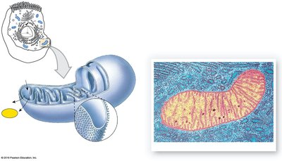
Mitochondria: Double-membraned organelles producing ATP through aerobic respiration.
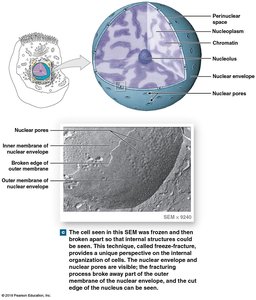
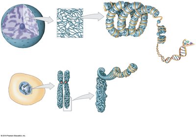
Nucleus: Contains DNA, nucleoli, and is surrounded by a nuclear envelope with pores.
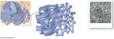
Endoplasmic Reticulum (ER): Network of membranes; rough ER (with ribosomes) synthesizes proteins, smooth ER synthesizes lipids and detoxifies chemicals.
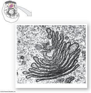
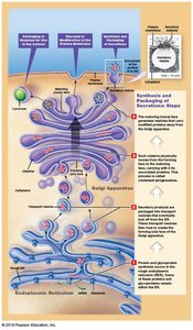
Golgi Apparatus: Modifies, sorts, and packages proteins and lipids for secretion or delivery to other organelles.
Lysosomes: Contain digestive enzymes for breakdown of waste and cellular debris.
Peroxisomes: Contain catalase to break down hydrogen peroxide; abundant in liver cells.
Intercellular Attachment
Cell Junctions
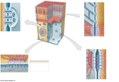
Cells form attachments with each other using cell adhesion molecules (CAMs) and intercellular cement (proteoglycans). Major types of cell junctions include:
Gap junctions: Allow free diffusion of ions and small molecules between cells.
Tight junctions (zonula occludens): Prevent passage of substances between cells.
Desmosomes (spot and hemidesmosomes): Provide strong adhesion between cells or to the extracellular matrix.
The Cell Life Cycle
Phases of the Cell Cycle
The cell life cycle includes interphase (cell growth and DNA replication) and mitosis (nuclear division), followed by cytokinesis (cytoplasmic division).
Interphase: Includes G0 (resting), G1 (organelle duplication), S (DNA synthesis), and G2 (protein synthesis).
Mitosis: Prophase, metaphase, anaphase, telophase
Cytokinesis: Division of the cytoplasm, resulting in two daughter cells.
DNA Replication: Occurs during S phase; DNA polymerase and ligases synthesize new DNA strands, resulting in duplicated chromosomes.
Mitosis Stages:
Prophase: Chromosomes condense, spindle fibers form.
Metaphase: Chromosomes align at the metaphase plate.
Anaphase: Sister chromatids separate to opposite poles.
Telophase: Nuclear membranes reform, chromosomes decondense.
Cytokinesis: Cleavage furrow divides the cytoplasm, completing cell division.
Phase | Main Events |
|---|---|
G1 | Organelle duplication, protein synthesis |
S | DNA replication, histone synthesis |
G2 | Protein synthesis, preparation for mitosis |
Mitosis | Prophase, metaphase, anaphase, telophase |
Cytokinesis | Division of cytoplasm |
Additional info: The cell cycle is tightly regulated to ensure proper growth, development, and maintenance of tissues. Errors in cell cycle regulation can lead to diseases such as cancer.
