 Back
BackChapter 20: The Lymphatic System and Lymphoid Organs and Tissues - Study Guide
Study Guide - Smart Notes
 Tailored notes based on your materials, expanded with key definitions, examples, and context.
The Lymphatic System: Structure and Function
Overview of the Lymphatic System
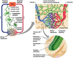
The lymphatic system is a network of vessels, organs, and tissues that plays a crucial role in fluid balance, immune defense, and fat absorption. It consists of lymphatic vessels, lymph (the fluid), lymph nodes, and lymphoid organs.
Lymphatic vessels collect excess interstitial fluid and return it to the bloodstream.
Lymph is the fluid transported within these vessels.
Lymph nodes filter lymph and house immune cells.
Lymphoid organs include the spleen, thymus, tonsils, and other tissues.
Functions of the Lymphatic System
The lymphatic system serves several essential functions:
Returns excess tissue fluid to the blood, maintaining fluid balance.
Returns leaked proteins to the blood.
Transports pathogens to lymph nodes for immune surveillance.
Absorbs fats from the intestine via specialized capillaries called lacteals.
Lymphatic Capillaries: Structure and Permeability
Structure and Unique Features
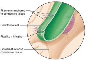
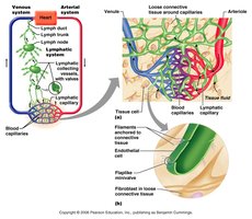
Lymphatic capillaries are the starting point of the lymphatic system. They are larger and more permeable than blood capillaries, allowing them to collect proteins, pathogens, and cell debris from interstitial fluid.
One-way minivalves: Endothelial cells overlap loosely, forming valves that open when interstitial fluid pressure increases.
Anchoring filaments: Collagen filaments attach capillaries to surrounding tissue, ensuring valves open with increased fluid volume.
Closed at one end: Unlike blood capillaries, lymphatic capillaries are blind-ended.
Permeability and Specialized Capillaries
Lymphatic capillaries are highly permeable, especially in inflamed tissues, allowing uptake of large molecules such as pathogens and cancer cells. In the small intestine, lacteals absorb digested fats, forming fatty lymph called chyle.
Lymph Flow and Major Vessels
Pathway of Lymph Flow
Lymph is generated from fluid that escapes blood capillaries and is collected by lymphatic capillaries. Its flow is facilitated by skeletal muscle contraction, respiratory movements, and valves.
Flow sequence: Capillaries → vessels → trunks → ducts
Lymphatic vessels anastomose and supply/drain lymph nodes.
Lymphatic Ducts
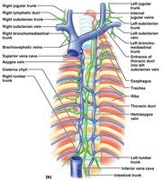
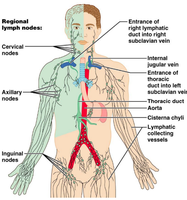
Right lymphatic duct: Drains lymph from the upper right side of the body.
Thoracic duct: Main collecting duct, drains most of the body, begins at the cisterna chyli.
Lymphoid Cells and Tissues
Lymphoid Cells
Lymphoid cells are found in lymphoid organs and tissues and include:
Lymphocytes: B cells (produce antibodies, memory cells) and T cells (cytotoxic, helper, suppressor, memory types).
Macrophages: Phagocytize foreign substances and activate T cells.
Dendritic cells: Capture antigens and activate T lymphocytes.
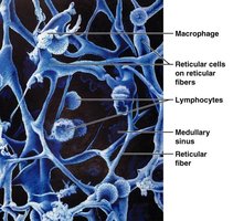
Reticular cells: Produce reticular fibers that form the stroma (supportive network).
Lymphoid Tissue
Most lymphoid organs are composed of reticular connective tissue, which houses lymphocytes and provides sites for their activation and proliferation.
Diffuse lymphatic tissue: Scattered reticular tissue in most organs.
Lymphoid follicles (nodules): Solid balls of reticular fibers and lymphoid cells, often with germinal centers.
Lymphoid Organs: Primary and Secondary
Classification of Lymphoid Organs
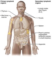
Primary lymphoid organs: Sites of lymphocyte maturation (red bone marrow for B cells, thymus for T cells).
Secondary lymphoid organs: Sites where mature lymphocytes encounter antigens (lymph nodes, spleen, MALTs).
The Thymus Gland
Structure and Function
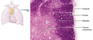
The thymus is a two-lobed organ located between the sternum and aorta. It is essential for T cell maturation and atrophies with age.
Outer cortex: Immature T cells proliferate and mature.
Inner medulla: Mature T cells enter the blood.
No follicles: Lacks B cells; functions strictly in T cell maturation.
Blood-thymus barrier: Prevents premature activation of T cells.
Lymph Nodes
Structure and Function
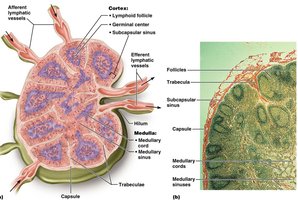
Lymph nodes are oval, bean-shaped structures that filter lymph and house immune cells. They are concentrated in regions such as the respiratory tree, GI tract, mammary glands, axillae, and groin.
Capsule: Fibrous connective tissue covering.
Trabeculae: Connective tissue strands extending inward.
Stroma: Supportive network of reticular fibers.
Cortex and Medulla
Cortex: Contains follicles with germinal centers (B cells) and deeper regions with T cells.
Medulla: Contains medullary cords with B and T lymphocytes.
Lymph Circulation in Nodes
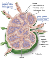
Lymph enters through afferent vessels, passes through sinuses, and exits via efferent vessels at the hilum. Fewer efferent vessels cause lymph to stagnate, allowing immune cells time to act. 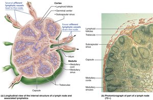
Lymph Sinuses
Lymph sinuses are large capillaries spanned by reticular fibers, where macrophages reside and phagocytize foreign matter. Only lymph nodes filter lymph. 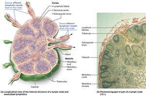
Clinical Aspects: Buboes, Metastasis, and Lymphadenopathy
Buboes and Metastasis
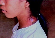
Buboes: Inflamed, swollen, and painful lymph nodes due to infection.
Metastasis: Cancer cells trapped in lymph nodes, usually not painful.
Lymphadenopathy
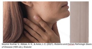
Lymphadenopathy: Enlarged lymph nodes, often due to increased drainage from infection or inflammation.
Lymphoid Organs: Spleen, Tonsils, and Peyer's Patches
Spleen
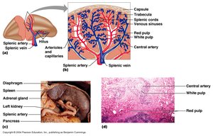
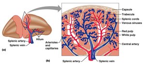
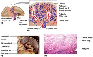
The spleen is the largest lymphoid organ, located between the stomach and diaphragm. It has a fibrous capsule and contains white and red pulp.
White pulp: Immune function, B cell maturation.
Red pulp: Blood cleansing, macrophage activity, storage of platelets and iron, site of fetal erythrocyte production.
Tonsils
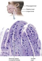
Tonsils are the simplest lymphoid organs, forming a ring around the throat entrance. They contain follicles with germinal centers and trap bacteria in tonsillar crypts.
Palatine tonsils: Largest and most frequently infected.
Peyer's Patches
Peyer's patches are large clusters of lymphoid follicles in the wall of the distal small intestine, providing immune surveillance. 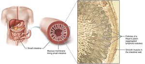
Diseases of the Lymphatic System
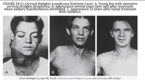
Hodgkin's Disease
Cause: Unknown, but Epstein-Barr virus is a contributing factor.
Curability: One of the most curable cancers.
Burkitt Lymphoma
Type: Non-Hodgkin's lymphoma, cancerous B cell.
African (endemic) type: Associated with Epstein-Barr virus, commonly involves the jaw.
American (sporadic) type: Not associated with EBV, extensive marrow replacement.
Immunodeficiency-associated type: Linked to HIV and immunosuppressive drugs.
Treatment: About half cured with extensive chemotherapy.
Summary Table: Major Lymphatic Structures and Functions
Structure | Main Function |
|---|---|
Lymphatic Capillaries | Collect interstitial fluid, absorb fats (lacteals) |
Lymph Nodes | Filter lymph, house immune cells |
Spleen | Immune response, blood cleansing, storage |
Thymus | T cell maturation |
Tonsils | Trap and destroy pathogens entering throat |
Peyer's Patches | Immune surveillance in intestine |
Additional info: The lymphatic system is essential for maintaining fluid balance, immune defense, and fat absorption. Its dysfunction can lead to edema, immune deficiencies, or increased risk of infection and cancer metastasis.
