 Back
BackChapter 20: The Lymphatic System and Immunity – Structured Study Notes
Study Guide - Smart Notes
 Tailored notes based on your materials, expanded with key definitions, examples, and context.
Module 20.1 Structure and Function of the Lymphatic System
Functions of the Lymphatic System
The lymphatic system is essential for maintaining fluid balance, absorbing dietary fats, and providing immune defense against pathogens.
Regulation of Fluid Volume: Not all fluid is reabsorbed by blood capillaries; excess fluid enters lymphatic vessels as lymph.
Absorption of Dietary Fats: Specialized lymphatic capillaries called lacteals absorb fats from the small intestine and transport them to the bloodstream.
Immune Functions: Lymphatic tissues filter pathogens and other harmful substances, preventing infection and disease.
Lymphatic Vessels and Circulation
Lymphatic vessels form a network that returns excess tissue fluid to the bloodstream and facilitate immune surveillance.
Lymphatic Capillaries: Weblike networks with closed ends, allowing one-way flow of lymph. Endothelial cell "flaps" permit entry of fluid and large solutes, including pathogens.
Lacteals: Specialized capillaries in the small intestine for fat absorption.
Lymph-Collecting Vessels: Larger, low-pressure vessels with valves to prevent backflow, located deep within muscle tissues.
Lymph Trunks: Nine major trunks collect lymph from specific body regions; the cisterna chyli is a collecting chamber for lower body lymph.
Lymphatic Ducts: Two main ducts drain lymph into the subclavian veins:
Thoracic Duct: Drains lymph from the left side and lower right side of the body into the left subclavian vein.
Right Lymphatic Duct: Drains lymph from the upper right side of the body into the right subclavian vein.
Lymphedema: Accumulation of excess fluid due to lymphatic obstruction or removal, often following surgery or immobility.
Lymphatic Tissues and Organs
Lymphatic tissues and organs are specialized for immune cell development, pathogen filtration, and immune response.
Lymphoid Cells:
Lymphocytes: B and T cells, central to adaptive immunity.
Phagocytes: Macrophages (mature monocytes) and dendritic cells (antigen-presenting leukocytes).
Reticular Cells: Provide structural support in organs like the spleen and lymph nodes.
Mucosa Associated Lymphatic Tissue (MALT): Clusters of B and T cells in mucous membranes (eyes, GI, oral, nasal, respiratory, urinary systems).
Tonsils: Capture and remove pathogens in the oral cavity.
Peyer's Patches: Lymphatic cell clusters in the small intestine, respond to pathogens.
Appendix: Contains lymphatic tissue, responds to waste and pathogens.
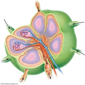
Lymph Nodes: Small, bean-shaped organs that filter lymph and stimulate immune responses. Located in axillary and cervical regions.
Cortex: Contains phagocytes and inactive B cells; deeper regions house T cells.
Medulla: Contains active B cells (plasma cells) that produce antibodies.
Flow: Lymph enters via afferent vessels, passes through cortex and medulla, and exits via efferent vessels.
Function: Prevents pathogens from entering the bloodstream.
Spleen: Largest lymphatic organ, filters blood.
Red Pulp: Contains macrophages, removes old red blood cells.
White Pulp: Contains leukocytes, filters pathogens.
Thymus: Located in the mediastinum, produces hormones for T cell maturation.
Module 20.2 Overview of the Immune System
Components and Lines of Defense
The immune system consists of leukocytes and proteins that defend against injury and pathogens. Defense is organized into three lines:
First Line: Skin and mucous membranes block pathogen entry.
Second Line: Non-specific cells and proteins (phagocytes, antimicrobial proteins).
Third Line: Specific cellular responses (adaptive immunity).
Types of Immunity
Innate (Nonspecific) Immunity: Rapid, general response to all pathogens; includes surface barriers, cells, and antimicrobial proteins.
Adaptive (Specific) Immunity: Slow, targeted response to specific antigens; involves immunological memory and two "arms":
Cell-mediated: T cells attack infected cells.
Antibody-mediated (Humoral): B cells produce antibodies.
Module 20.3 Innate Immunity: Internal Defenses
Cells of Innate Immunity
Innate immunity relies on phagocytic and nonphagocytic cells to eliminate pathogens.
Phagocytes: Macrophages (APCs), neutrophils, eosinophils, dendritic cells (APCs).
Nonphagocytic Cells: Natural Killer (NK) cells (cytotoxic), basophils (inflammatory mediators), mast cells (allergic response).
Antimicrobial Proteins
Complement: Liver-produced proteins activated via classical, lectin, or alternative pathways; effects include cell lysis, enhanced inflammation, virus neutralization, and phagocytosis.
Cytokines: Immune system proteins (tumor necrosis factor, interferon, interleukins) that regulate immune responses.
Inflammatory Response and Fever
Inflammation: Vasodilation, increased capillary permeability, pain, and chemotaxis recruit immune cells to injury sites.
Fever: Pyrogens act on the hypothalamus to raise body temperature, increasing metabolism and pathogen destruction.
Module 20.4 Adaptive Immunity: Cell-Mediated Immunity
Antigens and Classification
Antigens are substances recognized by T or B cells, triggering immune responses.
Immunogens: Foreign antigens from infected cells.
Self-antigens: Identify self cells to prevent autoimmunity.
Haptens: Small antigens that require a protein carrier to elicit a response (e.g., poison ivy).
Exogenous: Originates outside the cell, brought in by phagocytes.
Endogenous: Pathogens living inside cells.
T Cell Responses and MHC
T Cell Maturation: Occurs in the thymus; T cells develop specific receptors, self-tolerance, and immunocompetence.
Major Histocompatibility Complex (MHC): Glycoproteins on cell surfaces present antigens to T cells.
Class I MHC: Found on nucleated cells, present endogenous antigens to cytotoxic T cells.
Class II MHC: Found on APCs, present exogenous antigens to helper T cells.
T Cell Activation and Effects
Activation: Dendritic cells present antigens; T cells bind and undergo clonal selection and differentiation.
Helper T (TH) Cells: Secrete cytokines, stimulate macrophages, activate TC cells, and stimulate B cells.
Cytotoxic (TC) Cells: Destroy infected cells by releasing perforin and enzymes.
Transplantation and Rejection
Autograft: Self tissue, accepted.
Allograft: Tissue from another person, may be rejected if MHC genes do not match; immunosuppressive therapy is used to prevent rejection.
Module 20.5 Adaptive Immunity: Antibody-Mediated Immunity
B Cell Maturation and Activation
B cells mature in the bone marrow, enter circulation, and reside in lymphatic organs.
Self-reactive B cells: Destroyed to prevent autoimmunity.
Naïve B cell clones: Complete maturation and await activation.
Phase 1: B Cell Activation, Clonal Selection, and Differentiation
B cell binds antigen, processes it, and presents it on MHC molecules.
Helper T cells secrete cytokines to activate B cells.
B cells divide, differentiate into plasma cells (secrete antibodies) and memory B cells (long-lived).
Phase 2: Antibodies and Their Effects
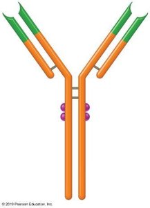
Antibodies are proteins produced by plasma cells that neutralize pathogens and facilitate their removal.
Structure:
Constant Region: Base for structural identification.
Variable Region: Unique, reflects antigen specificity.
Antigen Binding Sites: Bind to antigens.
Classes:
IgG: Most common, crosses placenta.
IgA: Present in secretions (saliva, mucus, tears, breast milk).
IgM: Largest, first responder.
IgE: Binds to cells with parasitic or environmental pathogens; involved in allergies.
IgD: Found on B cells, not secreted.
Functions: Neutralization, agglutination, complement activation, opsonization, stimulation of inflammation.
Phase 3: Immunological Memory
Primary Response: Slow, IgM produced first; peak in 14 days.
Secondary Response: Faster, IgG produced; peak in 3-5 days, lasts longer.
Vaccinations: Expose body to antigens, stimulate antibody production and memory cell formation.
Active Immunity: Body produces antibodies (infection or vaccination); memory cells formed.
Passive Immunity: Antibodies administered (placenta, breast milk, injections); no memory cells formed.
Module 20.7 Disorders of the Immune System
Hypersensitivity Disorders
Type I: Immediate Hypersensitivity: Allergens bind to B cells, plasma cells secrete IgE, mast cells sensitized; subsequent exposures trigger inflammation and possible anaphylactic shock.
Immunodeficiency Disorders
SCID: Failure to develop immune response; cells or antibodies not properly formed.
AIDS: Viral infection targets helper T cells, impairs antibody production.
Autoimmune Disorders
Multiple Sclerosis: Attacks myelin sheath, impairs nerve transmission.
Rheumatoid Arthritis: Autoantibodies attack joints.
Type I Diabetes Mellitus: Autoantibodies attack pancreas, halt insulin secretion.
Additional info: Both stress and aging diminish immune response.
