 Back
BackChapter 20: The Lymphatic System – Structure, Function, and Clinical Significance
Study Guide - Smart Notes
 Tailored notes based on your materials, expanded with key definitions, examples, and context.
The Lymphatic System: Overview
General Structure and Function
The lymphatic system is a vital component of human anatomy and physiology, consisting of a network of vessels and lymphoid organs. It plays essential roles in fluid balance, immune defense, and fat absorption.
Lymphatic Vessels: Transport lymph, a fluid derived from interstitial fluid, through capillaries, vessels, trunks, and ducts.
Lymphoid Tissues and Organs: Scattered throughout the body, these structures filter lymph and house immune cells.
Main Functions:
Returns interstitial fluid and leaked plasma proteins to the blood
Absorbs fats from the small intestine
Filters and destroys foreign antigens
Lymph: Interstitial fluid that enters lymphatic vessels, containing proteins, microbes, and sometimes cancer cells. About 3 liters are returned to the blood daily.
Movement: Lymph relies on skeletal and respiratory pumps for movement.
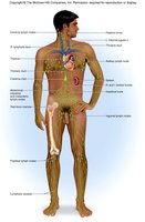
Lymphatic Capillaries
Structure and Permeability
Lymphatic capillaries are similar to blood capillaries but are more permeable due to specialized endothelial minivalves.
Minivalves: Act as one-way gates, allowing interstitial fluid to enter but preventing its escape.
One-way Flow: Ensures lymph moves in a single direction.
Role in Inflammation: Capillaries absorb cell debris, pathogens, and cancer cells during inflammation.
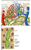
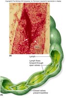
Lymphatic Collecting Vessels, Trunks, and Ducts
Organization and Drainage
Lymphatic vessels progress from capillaries to collecting vessels, trunks, and ducts, each with distinct structural features.
Collecting Vessels: Thinner walls and more internal valves than veins; travel alongside arteries and veins.
Trunks: Formed by the union of collecting vessels; major trunks include lumbar, intercostal, bronchiomediastinal, subclavian, jugular, and intestinal trunks.
Ducts:
Right Lymphatic Duct: Drains the right upper arm and right side of head and thorax.
Thoracic Duct: Arises from the cisterna chyli and drains the rest of the body.
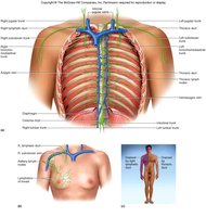
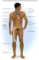
Clinical Significance: Blockage of Lymphatic Vessels
Elephantiasis and Lymphedema
Blockage of lymphatic vessels can lead to severe swelling known as edema.
Elephantiasis: Caused by parasitic worms clogging lymph vessels, resulting in massive swelling.
Lymphedema: General term for swelling due to lymphatic obstruction.


Lymphoid Tissue and Organs
Types and Functions
Lymphoid tissues and organs are specialized sites for immune cell development and function.
Diffuse Lymphatic Tissue: Scattered reticular tissue with macrophages and lymphocytes; includes MALT (mucosa-associated lymphatic tissue).
Lymphatic Nodules: Solid, spherical bodies with germinal centers of maturing B cells; examples include Peyer's Patches, tonsils, and appendix.
Lymphatic Organs: Well-defined anatomical sites with connective tissue capsules; examples are bone marrow, thymus, spleen, and lymph nodes.
Peyer's Patches
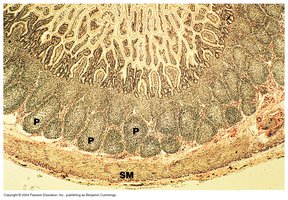
Peyer's Patches are clusters of lymphoid tissue in the small intestine, especially the ileum, and are similar to structures in the appendix.
Function: Destroy bacteria and prevent them from breaching the intestinal wall.
Tonsils
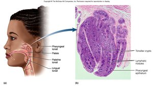
Tonsils are lymphatic nodules located in the oro/nasopharynx, filtering air and removing foreign materials.
Types:
Pharyngeal (adenoids): In nasopharynx near auditory tube
Palatine: On either side of oral cavity
Lingual: At the root of the tongue
Red Bone Marrow
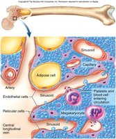
Red bone marrow is a primary lymphoid organ responsible for the production and maturation of B and T lymphocytes.
Location: In children, found in the medullary cavity of long bones; in adults, in the axial skeleton and proximal heads of humerus and femur.
Function: Contains sinusoids with macrophages that destroy malformed blood cells.
Thymus
The thymus is a primary lymphoid organ that secretes hormones (thymosin) for T lymphocyte maturation.
Development: Size varies with age; stops growing during adolescence and gradually atrophies.
Importance: Essential for the development of the immune system.
Secondary Lymphoid Organs
Secondary lymphoid organs are sites where lymphocytes encounter pathogens and initiate immune responses.
Includes: Lymph nodes and spleen.
Lymphatic Organ: Lymph Nodes
Structure and Function
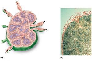
Lymph nodes are aggregations found near the body surface in various regions.
Functions: Filtration and immune system activation.
Cortex: Contains follicles with germinal centers rich in maturing B cells.
Deep Cortex: Houses T cells in transit.
Swelling: Occurs with infection or cancer.
Lymphatic Organ: Spleen
Structure and Functions
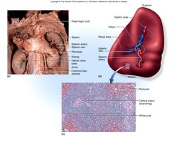
The spleen is the largest lymphoid organ, with multiple roles in immunity and blood maintenance.
Functions:
Site of B lymphocyte production
Cleanses blood
Stores products of RBC breakdown
Salvages and stores iron
Site of fetal erythrocyte production (ceases after birth)
Stores blood platelets
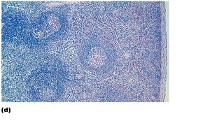
Structure: Surrounded by a fibrous capsule, contains trabeculae, lymphocytes, macrophages, and erythrocytes.
White Pulp: Area with lymphocytes involved in immune functions.
Red Pulp: Area concerned with disposing of worn-out RBCs and bloodborne pathogens.
Summary Table: Major Lymphatic Structures and Functions
Structure | Main Function | Location |
|---|---|---|
Lymphatic Capillaries | Absorb interstitial fluid, debris, pathogens | Throughout tissues |
Lymphatic Vessels | Transport lymph | Alongside veins/arteries |
Lymphatic Trunks/Ducts | Drain lymph from regions | Thoracic, right upper body |
Lymph Nodes | Filtration, immune activation | Body surface regions |
Spleen | Blood cleansing, immune response | Left upper abdomen |
Red Bone Marrow | Blood cell production | Axial skeleton, long bones |
Thymus | T cell maturation | Superior mediastinum |
Peyer's Patches | Bacterial defense | Ileum, appendix |
Tonsils | Air filtration | Oro/nasopharynx |
