 Back
BackChapter 22: The Respiratory System – ANP College Study Notes
Study Guide - Smart Notes
 Tailored notes based on your materials, expanded with key definitions, examples, and context.
Chapter 22: The Respiratory System
Overview and Functions of the Respiratory System
The respiratory system is essential for gas exchange, supplying oxygen to the body and removing carbon dioxide. It also plays roles in acid-base balance, olfaction, speech, and straining actions such as coughing.
Primary Functions: Intake of oxygen, expulsion of carbon dioxide, maintenance of acid-base balance.
Secondary Functions: Olfaction, speech, straining (e.g., coughing).
Relationship Between Respiratory and Cardiovascular Systems
The respiratory and cardiovascular systems work together to deliver oxygen to tissues and remove carbon dioxide. Gas exchange occurs in the lungs and systemic capillaries.
Conducting Zone: Includes respiratory muscles and airways that transport air.
Respiratory Zone: Includes pulmonary capillaries where gas exchange occurs.
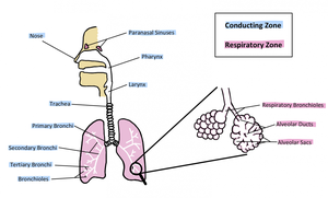
Divisions of the Respiratory System
The respiratory system is divided by location and function into upper and lower tracts, and conducting and respiratory zones.
Upper Respiratory Tract: Nasal/oral cavity through pharynx; warms, humidifies, and filters air.
Lower Respiratory Tract: Larynx to alveoli; site of gas exchange.
Conducting Zone: Nose to terminal bronchioles; not directly involved in gas exchange.
Respiratory Zone: Respiratory bronchioles to alveoli; directly involved in gas exchange.
22.1 Upper Respiratory System
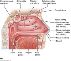
Nasal Cavity and Associated Structures
The nasal cavity is the first portion of the conducting zone, responsible for filtering, warming, and humidifying incoming air. It contains conchae (turbinates) that increase surface area and cause turbulence for better filtration.
Roof: Sphenoid, ethmoid, frontal, and nasal bones.
Lateral Walls: Superior, middle, and inferior nasal conchae.
Nasal Septum: Ethmoid bone, vomer, septal cartilage.
Hard Palate: Maxilla (anterior ¾), palatine bone (posterior ¼).
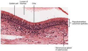
Paranasal Sinuses and Respiratory Epithelium
Paranasal sinuses lighten the skull and add resonance to the voice. The respiratory epithelium contains cilia and goblet cells that move mucus and debris toward the throat.
Sinuses: Located in frontal, ethmoid, sphenoid, and maxilla.
Respiratory Epithelium: Pseudostratified columnar tissue with goblet cells and cilia.
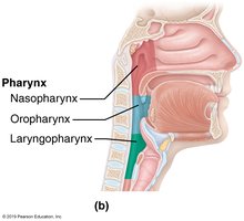
Pharynx
The pharynx is divided into three regions: nasopharynx (air only), oropharynx (air, food, liquid), and laryngopharynx (air, food, liquid). It contains tonsils and is involved in swallowing and airway protection.
Nasopharynx: Contains adenoids, auditory tube openings.
Oropharynx: Contains lingual and palatine tonsils.
Laryngopharynx: Opens into larynx and esophagus; epiglottis covers larynx during swallowing.
22.2 Lower Respiratory System
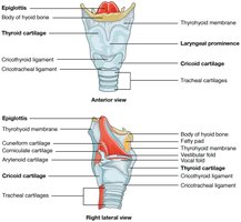
Larynx
The larynx marks the beginning of the lower respiratory tract, connecting the pharynx to the trachea and producing sound. It is formed by three major cartilages: epiglottis, thyroid, and cricoid.
Epiglottis: Covers airway during swallowing.
Thyroid Cartilage: Forms the "Adam's apple."
Cricoid Cartilage: Only full ring of cartilage.
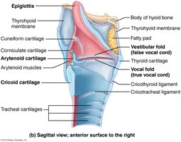
Glottis and Vocal Cords
The glottis includes the true vocal cords, which oscillate to produce sound. False vocal cords (vestibular folds) are found laterally.
True Vocal Cords: Produce sound.
False Vocal Cords: Vestibular folds.
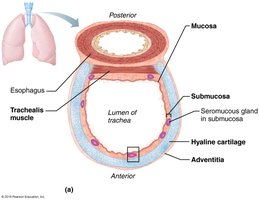
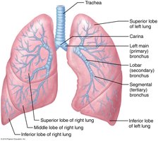
Trachea and Bronchi
The trachea is supported by C-shaped hyaline cartilage rings and contains the trachealis muscle. The carina is a cartilaginous ridge where the primary bronchi branch.
Trachea: C-shaped cartilage rings, trachealis muscle.
Carina: Site of bronchial bifurcation; sensitive to induce coughing.
Bronchi: Primary (right and left), secondary (lobar), tertiary (segmental), bronchioles.
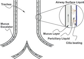
Mucociliary Escalator
The mucociliary escalator is a defense mechanism that clears debris from the respiratory tract by moving mucus upward toward the pharynx.
Cilia: Beat rhythmically to move mucus.
Goblet Cells: Produce mucus.
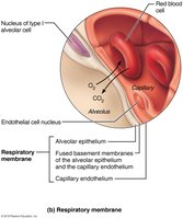
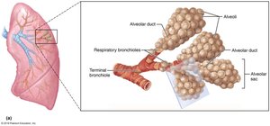
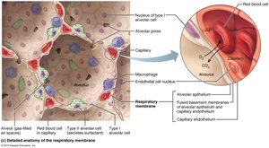
Respiratory Zone and Alveoli
The respiratory zone includes respiratory bronchioles and alveoli, where gas exchange occurs. Alveoli are lined with simple squamous epithelium and closely associated with capillaries.
Type 1 Alveolar Cells: Squamous epithelial cells for gas exchange.
Type 2 Alveolar Cells: Produce surfactant to reduce surface tension and prevent alveolar collapse.
Alveolar Macrophages: Consume debris and invaders.
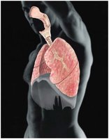
22.3 Lungs and Pleura
Lung Anatomy and Pleural Membranes
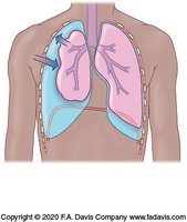
Each lung occupies its own pleural cavity and is encased in pleural membranes. The pleurae reduce friction, compartmentalize, and help inflate the lungs.
Visceral Pleura: Covers the lung surface.
Parietal Pleura: Lines the thoracic cavity.
Pleural Cavity: Contains serous fluid for lubrication.
22.4 Volume, Pressure, and Air Movement
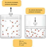
Boyle’s Law and Air Movement
Boyle’s Law states that pressure and volume are inversely related. Air moves from areas of high pressure to low pressure.
Equation:
Air Movement: Driven by pressure differences.
Breathing Muscles
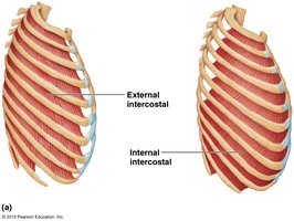
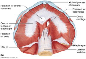
The diaphragm and intercostal muscles are essential for breathing. The diaphragm is the prime mover of inspiration, innervated by the phrenic nerves (C3, C4, C5).
External Intercostals: Elevate rib cage for inspiration.
Internal Intercostals: Depress rib cage for forced expiration.
Expiration at Rest: Passive process due to elastic recoil.
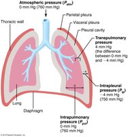
Pressure Relationships in Breathing
Atmospheric and intrapulmonary pressures equalize during breathing. Intrapleural pressure is negative, helping inflate the lungs.
Atmospheric Pressure: Pressure of air around us (760 mm Hg).
Intrapulmonary Pressure: Pressure within the lungs.
Intrapleural Pressure: Negative pressure in pleural space (~ -4 mm Hg).
Inspiration and Expiration
Inspiration is an active process involving diaphragm contraction and rib elevation, increasing thoracic volume and decreasing pressure. Expiration is passive at rest, with diaphragm relaxation and elastic recoil.
Inspiration: Diaphragm contracts, external intercostals lift ribs.
Expiration: Diaphragm relaxes, rib cage recoils.
Forced Expiration: Abdominal muscles contract, internal intercostals pull ribs down.
22.5 Spirometry and Respiratory Volumes
Respiratory Volumes and Capacities
Spirometry measures respiratory volumes and capacities, which are important for assessing lung function.
Tidal Volume (TV): Amount of air moved in and out during normal breathing (~500 mL).
Inspiratory Reserve Volume (IRV): Extra air inhaled beyond TV.
Expiratory Reserve Volume (ERV): Extra air exhaled beyond TV.
Residual Volume (RV): Air remaining after full exhalation (~1200 mL).
Obstructive vs. Restrictive Pulmonary Diseases
Obstructive diseases (e.g., COPD) increase airway resistance and lung volumes, while restrictive diseases (e.g., tuberculosis) limit lung expansion and decrease volumes.
Obstructive: Increased TLC and RV, decreased FEV1.
Restrictive: Decreased TLC and RV, FEV1 may be normal percentage.
Dead Space
Dead space refers to areas where air does not participate in gas exchange.
Anatomical Dead Space: Conducting zones (~150 mL).
Alveolar Dead Space: Alveoli not perfused or obstructed.
22.6 Gas Exchange by Diffusion
Dalton’s Law
Dalton’s Law states that the total pressure of a gas mixture is the sum of the partial pressures of its components.
Equation:
Henry’s Law
Henry’s Law states that gas diffuses into liquid in proportion to its partial pressure and solubility.
Clinical Application: Hyperbaric oxygen chambers increase O2 absorption.
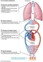
Internal and External Respiration
Gas exchange occurs in the alveoli (external respiration) and tissues (internal respiration) based on partial pressure gradients.
External Respiration: O2 diffuses from air to blood; CO2 from blood to air.
Internal Respiration: O2 diffuses from blood to tissues; CO2 from tissues to blood.
22.7 Oxygen and Carbon Dioxide Transport
Oxyhemoglobin Dissociation Curve
The curve shows how hemoglobin's affinity for oxygen changes with partial pressure. It is S-shaped due to conformational changes in hemoglobin.
Shift to Left: Hemoglobin holds O2 more tightly (decreased temperature, CO2, increased pH).
Shift to Right: Hemoglobin releases O2 more easily (increased temperature, CO2, decreased pH).
Carbon Dioxide Transport
CO2 travels in blood bound to hemoglobin, dissolved in plasma, or as bicarbonate ions.
Bound to Hemoglobin: ~20%
Dissolved in Plasma: ~10%
As Bicarbonate: ~70%
22.8 Respiratory Control
Brain Stem Centers and Chemoreceptors
Breathing is controlled by the medulla and pons, which receive input from chemoreceptors and higher brain centers.
Medulla: Generates and modifies breathing rhythm.
Pons: Smooths respiratory pattern.
Autonomic Innervation: Sympathetic dilates bronchioles; parasympathetic constricts.
Control of Respiration: Carbon Dioxide
CO2 is the most powerful stimulant for breathing. Accumulation causes blood acidity, stimulating chemoreceptors to increase respiration.
Hypercapnia: High CO2, low pH, increased breathing rate.
Hypocapnia: Low CO2, can cause apnea.
Special Topic: Acid-Base Balance
Blood pH Regulation
Normal blood pH is 7.35–7.45. The respiratory system, chemical buffers, and renal system regulate acid-base balance.
Alkalosis: Blood less acidic, pH rises.
Acidosis: Blood more acidic, pH drops.
Buffers: Chemical, respiratory, and renal systems.
References: Marieb & Hoehn, Marieb & Smith, Thompson, Gordon et al., OpenStax, Etymonline.com.
