 Back
BackChapter 22: The Respiratory System – Functional Anatomy and Physiology
Study Guide - Smart Notes
 Tailored notes based on your materials, expanded with key definitions, examples, and context.
Overview of the Respiratory System
The respiratory system is essential for supplying cells with oxygen and eliminating carbon dioxide. It is divided into two main parts: Functional Anatomy and Respiratory Physiology. This chapter explores the structure, function, and physiological mechanisms of the respiratory system, as well as its developmental aspects and common diseases.
Part 1: Functional Anatomy
22.1 The Upper Respiratory System
The upper respiratory system includes the nose, paranasal sinuses, and pharynx. These structures warm, humidify, and filter incoming air, providing protection and facilitating respiration.
Nose: Provides an airway, moistens and warms air, filters particles, and houses olfactory receptors.
Paranasal Sinuses: Lighten the skull, warm and moisten air, and produce mucus.
Pharynx: Connects the nasal cavity and mouth to the larynx and esophagus; divided into nasopharynx, oropharynx, and laryngopharynx.
Protective Mechanisms: Include mucus production, lysozyme, defensins, and tonsils that trap pathogens.
22.2 The Lower Respiratory System
The lower respiratory system consists of the larynx, trachea, bronchi, and alveoli. It is divided into conducting and respiratory zones, facilitating the passage and exchange of gases.
Larynx: Maintains an open airway, routes food and air, and produces sound.
Trachea: Supported by cartilaginous rings, lined with ciliated epithelium to propel mucus.
Bronchi and Bronchioles: Branch into smaller airways, with structural changes and increased smooth muscle.
Alveoli: Site of gas exchange, surrounded by capillaries and elastic fibers; type I cells form the respiratory membrane, type II cells secrete surfactant.
22.3 The Lungs and Pleurae
Each lung occupies its own pleural cavity and is divided into lobes and segments. The pleurae are double-layered serous membranes that lubricate and protect the lungs.
Lobes and Segments: Right lung has three lobes, left lung has two; each lobe contains bronchopulmonary segments.
Pleurae: Parietal pleura lines the thoracic wall; visceral pleura covers the lung surface; pleural fluid reduces friction.
Blood Supply: Pulmonary arteries oxygenate blood; bronchial arteries supply lung tissue.
Part 2: Respiratory Physiology
22.4 Pulmonary Ventilation
Volume changes in the thoracic cavity cause pressure changes, leading to air movement. Boyle’s law governs the relationship between pressure and volume.
Intrapulmonary Pressure: Pressure within alveoli; equalizes with atmospheric pressure.
Intrapleural Pressure: Pressure within pleural cavity; always lower than intrapulmonary pressure.
Transpulmonary Pressure: Difference between intrapulmonary and intrapleural pressure; determines lung volume.
Physical Factors: Airway resistance, alveolar surface tension, and lung compliance affect ventilation.
Boyle’s Law:
22.5 Respiratory Volumes and Capacities
Respiratory volumes and capacities are measured to assess ventilation and lung function.
Tidal Volume (TV): Amount of air moved per breath (500 ml).
Inspiratory Reserve Volume (IRV): Additional air inspired (2100–3200 ml).
Expiratory Reserve Volume (ERV): Additional air expired (1000–1200 ml).
Residual Volume (RV): Air remaining after maximal expiration (1200 ml).
Capacities: Sums of volumes (IC, FRC, VC, TLC).
Anatomical Dead Space: Conducting zone volume not involved in gas exchange (150 ml).
Pulmonary Function Tests: Distinguish obstructive vs. restrictive disorders.
22.6 Gas Exchange: Diffusion and Laws
Gases exchange by diffusion between blood, lungs, and tissues, governed by Dalton’s and Henry’s laws.
Dalton’s Law: Total pressure is the sum of partial pressures of each gas.
Henry’s Law: Gas dissolves in liquid in proportion to its partial pressure.
External Respiration: Oxygen uptake and carbon dioxide unloading in lungs.
Internal Respiration: Oxygen delivery and carbon dioxide removal in tissues.
Ventilation-Perfusion Coupling: Matches air flow and blood flow for optimal gas exchange.
22.7 Oxygen and Carbon Dioxide Transport
Oxygen is transported by hemoglobin; carbon dioxide is transported dissolved in plasma, bound to hemoglobin, or as bicarbonate.
Oxygen Transport: 98.5% bound to hemoglobin, 1.5% dissolved in plasma.
Factors Affecting Oxygen Loading/Unloading: Temperature, pH, BPG, and partial pressures.
Carbon Dioxide Transport: 7–10% dissolved, 20% bound to hemoglobin, 70% as bicarbonate.
Carbonic Acid–Bicarbonate Buffer:
Hypoxia: Inadequate oxygen delivery; classified as anemic, ischemic, histotoxic, hypoxemic, or due to CO poisoning.
22.8 Neural Control of Respiration
Respiratory centers in the brain stem regulate breathing, influenced by chemoreceptors, higher brain centers, and reflexes.
Medulla: Ventral and dorsal respiratory groups generate and integrate breathing rhythm.
Pons: Pontine respiratory group modifies rhythm and prevents over-inflation.
Chemoreceptors: Monitor CO2, O2, and pH; central (medulla) and peripheral (aortic/carotid bodies).
Higher Brain Centers: Hypothalamus, limbic system, and cortex influence breathing.
Reflexes: Pulmonary irritant and inflation (Hering-Breuer) reflexes protect airways and lungs.
22.9 Respiratory Adjustments: Exercise and High Altitude
Exercise and high altitude require respiratory adjustments to meet oxygen demands and adapt to environmental changes.
Hyperpnea: Increased depth and rate of breathing during exercise.
Hyperventilation: Excessive ventilation leading to low CO2 and alkalosis.
Acclimatization: Long-term adaptation to high altitude includes increased ventilation and erythropoietin production.
22.10 Respiratory Diseases
Respiratory diseases are major causes of disability and death, including COPD, asthma, tuberculosis, and lung cancer.
COPD: Includes emphysema (damaged alveoli) and chronic bronchitis (excess mucus and inflammation).
Asthma: Inflammation and constriction of airways.
Tuberculosis: Infectious disease caused by Mycobacterium tuberculosis.
Lung Cancer: Most common malignancy; types include adenocarcinoma, squamous cell carcinoma, and small cell carcinoma.
Developmental Aspects of the Respiratory System
The respiratory system develops from embryonic structures and undergoes changes throughout life. Aging affects elasticity, mucus production, and immune function.
Embryonic Development: Olfactory placodes form nasal cavities; laryngotracheal bud forms lower respiratory organs.
Fetal Lungs: Filled with fluid; at birth, fluid drains and breathing begins.
Aging: Decreased elasticity, increased rigidity, reduced oxygen use, and diminished immune defenses.
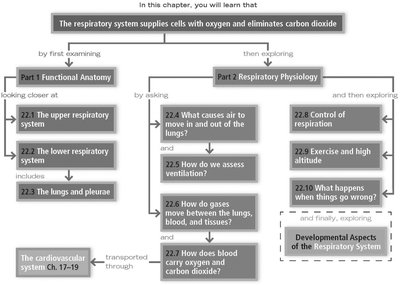
Summary Table: Major Divisions and Functions of the Respiratory System
Division | Main Structures | Primary Functions |
|---|---|---|
Upper Respiratory System | Nose, Paranasal Sinuses, Pharynx | Warm, humidify, filter air; protect against pathogens |
Lower Respiratory System | Larynx, Trachea, Bronchi, Alveoli | Conduct air, produce sound, gas exchange |
Lungs & Pleurae | Lobes, Segments, Pleural Membranes | Gas exchange, protection, lubrication |
Respiratory Physiology | Muscles, Membranes, Blood Vessels | Ventilation, gas transport, neural control |
Additional info:
Recent discoveries highlight new lung functions (platelet production), gender differences in asthma, and dietary effects on lung repair.
Cross-references to other chapters provide context for related anatomical and physiological concepts.
