Back
BackChapter 22: The Respiratory System – Structure, Function, and Clinical Relevance
Study Guide - Smart Notes
 Tailored notes based on your materials, expanded with key definitions, examples, and context.
The Respiratory System: Overview
Introduction to the Respiratory System
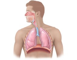
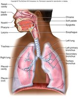
The respiratory system is essential for gas exchange, supplying oxygen to the body and removing carbon dioxide. It consists of anatomical structures that conduct air and facilitate gas exchange, divided into the conducting and respiratory zones.
Conducting Zone: Transports, cleanses, humidifies, and warms incoming air. Includes the nose, nasal cavity, pharynx, larynx, trachea, bronchi, and bronchioles.
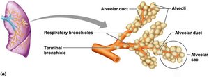
Respiratory Zone: Site of gas exchange, consisting of respiratory bronchioles, alveolar ducts, and alveoli.
Anatomy of the Upper Respiratory Tract
Pharynx: Regions and Functions
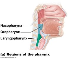
The pharynx is a muscular tube that serves as a passageway for both air and food, divided into three regions:
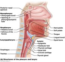
Nasopharynx: Lined with pseudostratified ciliated columnar epithelium; humidifies and warms air, propels mucus downward, and is closed off during swallowing.
Oropharynx: Lined with stratified squamous epithelium; common passageway for food and air, contains palatine and lingual tonsils.
Laryngopharynx: Also lined with stratified squamous epithelium; serves as a common passageway and diverges into the larynx (air) and esophagus (food).
Larynx and Trachea
Larynx (Voice Box): Structure and Function
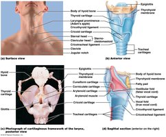
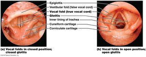
The larynx routes air and food into proper channels and is responsible for voice production. It is composed of several cartilages and contains the vocal cords.
Cartilages: Thyroid (Adam’s apple), cricoid, arytenoid, cuneiform, corniculate, and epiglottis.
Vocal Ligaments: True vocal cords (vocal folds) and false vocal cords (vestibular folds); the glottis is the medial opening.
Movements of the Vocal Cords
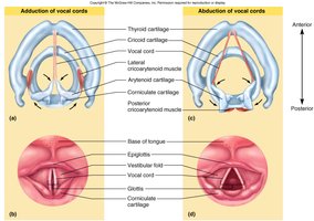
The vocal cords can be adducted (closed) or abducted (open) to control airflow and sound production.
Trachea
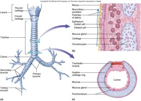
The trachea is a flexible tube lined with pseudostratified ciliated columnar epithelium and reinforced by C-shaped hyaline cartilage rings. It branches into the primary bronchi at the carina.
Bronchial Tree and Respiratory Zone
Bronchi and Bronchioles
The bronchial tree consists of branching airways that decrease in size and change in structure as they penetrate deeper into the lungs.
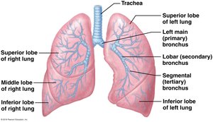
Primary Bronchi: Enter each lung.
Secondary (Lobar) Bronchi: Serve each lobe.
Tertiary (Segmental) Bronchi: Serve bronchopulmonary segments.
Bronchioles: Lack cartilage, have smooth muscle, and are the site of asthma attacks.
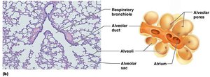
Respiratory Zone: Alveoli
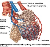
The respiratory zone includes respiratory bronchioles and alveoli, where gas exchange occurs. Alveoli are made of simple squamous epithelium and are surrounded by capillaries and elastic fibers.
Type I Alveolar Cells: Form the alveolar wall for gas diffusion.
Type II Alveolar Cells: Produce surfactant to reduce surface tension.
Alveolar Macrophages (Dust Cells): Remove pathogens and debris.
Respiratory Membrane
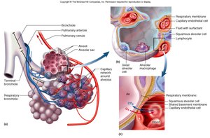
The respiratory membrane is the site of gas exchange, consisting of the alveolar and capillary walls, a thin interstitial space, and surfactant fluid. Factors affecting gas exchange include membrane thickness, surface area, partial pressure differences, and diffusion coefficient.
Lungs and Pleura
Lung Structure and Pleural Membranes
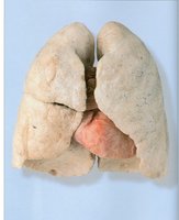
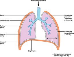
The lungs are divided into lobes and covered by a double-layered serous membrane (pleura). The parietal pleura lines the thoracic cavity, while the visceral pleura covers the lungs. Pleural fluid reduces friction and creates a pressure gradient.
Right Lung: Three lobes (superior, middle, inferior).
Left Lung: Two lobes (superior, inferior) and a cardiac notch.
Mechanics of Breathing (Ventilation)
Principles of Ventilation
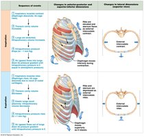
Breathing involves creating a pressure gradient so that air flows from high to low pressure. Boyle’s Law states that pressure and volume are inversely proportional: .
Inspiration: Air moves into the lungs as thoracic volume increases and pressure decreases.
Expiration: Air moves out as thoracic volume decreases and pressure increases.
Pressure Relationships
Key pressures include:
Intrapulmonary Pressure (Ppul): Pressure within alveoli.
Intrapleural Pressure (Pip): Pressure within pleural cavity (normally -4 mm Hg relative to atmospheric pressure).
Atmospheric Pressure (Patm): Pressure of the air outside the body (760 mm Hg at sea level).
Factors Preventing Lung Collapse
Surfactant: Reduces surface tension in alveoli, preventing collapse.
Negative Intrapleural Pressure: Maintains lung expansion.
Residual Volume: Air remaining after expiration keeps alveoli open.
Lung Compliance
Lung compliance refers to the ease with which the lungs can expand. It is diminished by fibrosis, airway obstruction, reduced surfactant, or decreased thoracic flexibility.
Respiratory Volumes and Capacities
Key Volumes
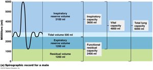
Tidal Volume (TV): Air moved in/out per breath (~500 ml).
Inspiratory Reserve Volume (IRV): Extra air inhaled after normal inspiration (~3100 ml).
Expiratory Reserve Volume (ERV): Extra air exhaled after normal expiration (~1200 ml).
Residual Volume (RV): Air remaining after maximal expiration (~1200 ml).
Key Capacities
Inspiratory Capacity (IC):
Functional Residual Capacity (FRC):
Vital Capacity (VC):
Total Lung Capacity (TLC): (about 6000 ml in males)
Minute and Alveolar Ventilation
Minute Respiratory Volume: (e.g., 500 ml × 12 breaths/min = 6000 ml/min)
Alveolar Ventilation Rate (AVR): (e.g., 12 × (500 – 150) = 4200 ml/min)
Gas Exchange and Transport
Partial Pressures and Gas Laws
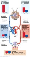
Dalton’s Law: The partial pressure of each gas in a mixture is proportional to its percentage. Gases move from higher to lower partial pressure.
Oxygen Transport
98.5% of O2 is bound to hemoglobin (Hb) as oxyhemoglobin (HbO2).
1.5% is dissolved in plasma.
Hemoglobin can bind up to 4 O2 molecules; saturation depends on PO2.
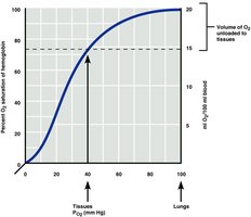
Hemoglobin-Oxygen Dissociation Curve
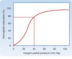
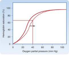
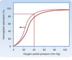
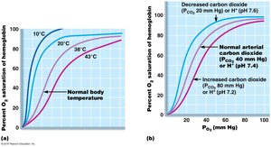
This curve shows the relationship between PO2 and hemoglobin saturation. It is influenced by pH, CO2, temperature, and BPG (2,3-bisphosphoglycerate).
Carbon Dioxide Transport
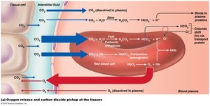
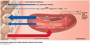
7–10% dissolved in plasma
20% bound to hemoglobin as carbaminohemoglobin
70% as bicarbonate ion (HCO3–) in plasma
CO2 + H2O H2CO3 H+ + HCO3–
Buffer Systems and pH Regulation
The carbonic acid–bicarbonate buffer system resists changes in blood pH. Hemoglobin also acts as a buffer by binding H+ ions (Bohr Effect).
Control of Respiration
Neural Control Centers
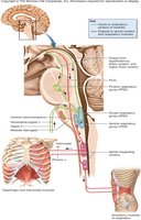
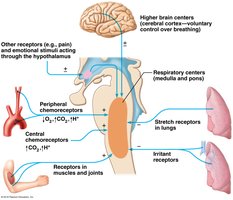
Medulla Oblongata: Ventral respiratory group (VRG) sets basic rhythm; dorsal respiratory group (DRG) integrates input.
Pons: Modifies and smooths respiratory rhythm.
Other Controls
Hering-Breuer Reflex: Prevents overinflation of lungs via stretch receptors.
Conscious Control: Voluntary control is limited; brain centers can override willful changes.
Pulmonary Irritants: Trigger constriction of air passages.
Chemical Regulation
CO2 is the primary regulator; increased CO2 stimulates increased ventilation.
O2 has less influence unless levels drop significantly.
Respiratory Diseases and Disorders
Common Diseases
Hypoxia: Inadequate O2 delivery due to various causes.
Carbon Monoxide Poisoning: CO binds to Hb with high affinity, displacing O2.
Hyperventilation: Decreases CO2, leading to alkalosis.
Acute Mountain Sickness: Low PO2 at high altitudes; body acclimates by increasing RBC production.
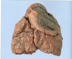
Tuberculosis: Bacterial infection causing lung fibrosis.
Asthma: Bronchospasm and airway inflammation.
Sleep Apnea: Periodic cessation of breathing during sleep.
Cystic Fibrosis: Genetic disorder causing thick mucus and infections.
Pulmonary Embolism: Blockage of pulmonary artery by a clot.
Pneumonia: Infection of the lungs by bacteria or viruses.
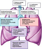
Chronic Obstructive Pulmonary Disease (COPD): Includes emphysema and chronic bronchitis; characterized by airflow obstruction and lung tissue damage.
Lung Cancer: Strongly associated with smoking; high mortality rate.