 Back
Chapter 22: The Respiratory System – Structure, Function, and Regulation
Study Guide - Smart Notes
 Tailored notes based on your materials, expanded with key definitions, examples, and context.
Respiratory System Overview
Processes of Respiration
The respiratory system supplies the body with oxygen and removes carbon dioxide through four distinct processes:
Pulmonary ventilation: Movement of air into and out of the lungs (breathing).
External respiration: Gas exchange between the blood and alveoli (air sacs of the lungs).
Transport of respiratory gases: Movement of gases to and from the lungs and tissue cells via the bloodstream.
Internal respiration: Gas exchange between blood and tissue cells at systemic capillaries.
Anatomy of the Respiratory System
Organs and Zones
The respiratory system consists of the nose, pharynx, larynx, trachea, bronchi, and alveoli. It is divided into two functional zones:
Conducting zone: Includes all respiratory passageways except the respiratory bronchioles, alveolar ducts, and alveoli. Its main functions are to purify, humidify, and warm incoming air.
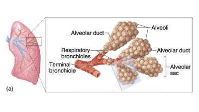
Respiratory zone: The site of actual gas exchange, consisting of respiratory bronchioles, alveolar ducts, and alveoli.
Functions of Major Respiratory Structures
Nose: Provides an airway, moistens and warms air, filters and cleanses air, serves as a resonating chamber for speech, and houses olfactory receptors. Mucus and cilia trap and move contaminants.
Pharynx: Connects the nasal cavity and mouth to the larynx and esophagus. It is divided into three regions:
Nasopharynx: Air passage only; uvula prevents food entry during swallowing.
Oropharynx: Passageway for food and air.
Laryngopharynx: Passageway for food and air; continuous with the esophagus.
Larynx (voice box): Maintains an open airway, routes air and food, and houses the vocal cords. The epiglottis acts as a switching mechanism during swallowing.
Trachea: Extends from the larynx and divides into two primary bronchi. Ciliated cells move mucus and debris upward toward the pharynx.
Bronchial Tree Structure and Function
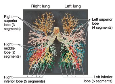
The bronchial tree consists of branching airways that conduct air from the trachea into the lungs:
Right and left primary bronchi enter the lungs at the hilus.
Each bronchus subdivides into secondary and tertiary bronchi, continuing for 23 orders of branching.
The smallest branches are called bronchioles.
Respiratory Membrane and Gas Exchange
The respiratory membrane is formed by the alveolar and capillary walls, allowing efficient gas exchange by simple diffusion. Key features include:
Alveoli are composed of a single layer of epithelium.
Surfactant produced by alveolar cells reduces surface tension, preventing alveolar collapse.
Alveolar macrophages defend against inhaled particles and pathogens.
Lungs and Pleural Coverings
Each lung is housed in its own pleural cavity and rests on the diaphragm. The lungs differ in size and shape due to the position of the heart:
Right lung: Three lobes (superior, middle, inferior).
Left lung: Two lobes (superior, inferior).
The pleura are serous membranes:
Visceral pleura: Covers the lung surface.
Parietal pleura: Lines the thoracic cavity.
Pleural fluid between the layers reduces friction during breathing.
Mechanics of Breathing
Inspiration and Expiration
Breathing involves two phases:
Inspiration: Diaphragm contracts, thoracic cavity volume increases, intrapulmonary pressure decreases, and air flows into the lungs.
Expiration: Usually passive; lung elasticity causes thoracic volume to decrease, intrapulmonary pressure increases, and air flows out. Forced expiration uses abdominal muscles.
Negative Intrapleural Pressure
The pressure within the pleural cavity (intrapleural pressure) is always negative relative to atmospheric and intrapulmonary pressures, preventing lung collapse. This is maintained by:
Adhesive force of pleural fluid.
Positive pressure within the lungs and atmospheric pressure on the thorax.
Opposing forces: lung recoil and alveolar surface tension pull lungs away from the thorax wall.
Physical Factors Influencing Pulmonary Ventilation
Volume changes in the thoracic cavity lead to pressure changes, driving airflow.
Inspiration depends on diaphragm contraction; expiration is usually passive.
Forced expiration involves abdominal muscles.
Role of Surfactant
Surfactant is a detergent-like substance produced by alveolar cells that reduces surface tension in the alveoli, making it easier to expand the lungs and preventing alveolar collapse.
Respiratory Volumes and Capacities
Tidal volume (TV): Air moved in and out during normal breathing.
Inspiratory reserve volume (IRV): Air that can be forcibly inhaled after a normal inspiration.
Expiratory reserve volume (ERV): Air that can be forcibly exhaled after a normal expiration.
Vital capacity (VC): Total amount of exchangeable air.
Gas Exchange and Transport
Factors Influencing Gas Exchange
Pressure gradients: Oxygen diffuses from alveoli (104 mm Hg) to blood (40 mm Hg); carbon dioxide diffuses in the opposite direction.
Respiratory membrane structure: Thin and large surface area enhances diffusion.
Ventilation-perfusion matching: Blood flow is directed to well-ventilated areas for efficient gas exchange.
Control of Respiration
Neural and Chemical Regulation
Medullary respiratory centers: Generate basic rhythm of breathing.
Pontine respiratory centers: Modify and smooth respiratory rhythm, especially during speech, sleep, and exercise.
Chemical factors:
CO2 levels are the most powerful stimulus for breathing.
Low O2 and low pH also stimulate respiration.
Other factors: Exercise and high altitude can increase respiratory rate.
Respiratory Disorders
Chronic obstructive pulmonary disease (COPD):
Emphysema: Permanent enlargement of alveoli, destruction of walls, loss of elasticity.
Chronic bronchitis: Chronic mucus production, inflammation, and fibrosis of lower respiratory mucosa.
Asthma: Reversible airway obstruction with episodes of coughing, wheezing, and chest tightness; caused by airway inflammation.
Tuberculosis: Infection by Mycobacterium tuberculosis; forms nodules (tubercles) in the lungs.
Lung cancer:
Squamous cell carcinoma: Originates in bronchial epithelium.
Adenocarcinoma: Originates in peripheral lung areas.
Small cell carcinoma: Originates in main bronchi.
