 Back
BackChapter 23: The Digestive System – Overview and Structure
Study Guide - Smart Notes
 Tailored notes based on your materials, expanded with key definitions, examples, and context.
Overview of the Digestive System
Main Groups of Digestive Organs
The digestive system consists of two main groups: the alimentary canal and accessory digestive organs. Together, these structures facilitate the breakdown and absorption of nutrients necessary for bodily function.
Alimentary Canal: Also known as the gastrointestinal (GI) tract, this continuous muscular tube extends from the mouth to the anus. It is technically outside the body because it is open to the external environment at both ends. Its primary functions are to digest food and absorb nutrients.
Accessory Digestive Organs: These include glands and other structures that lie outside the GI tract, such as the teeth, tongue, gallbladder, salivary glands, liver, and pancreas. They produce secretions that help break down food.
Alimentary Canal Organs: Mouth, pharynx, esophagus, stomach, small intestine, large intestine (leading to the anus).
Accessory Organs: Teeth, tongue, gallbladder, salivary glands, liver, pancreas.
Processing of Food: Essential Activities
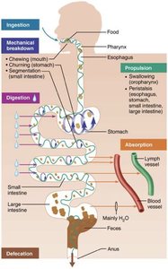
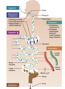
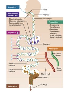
The digestive system acts as a "disassembly line," breaking down food into simpler components as it passes through. There are six essential activities:
Ingestion: Taking food into the digestive tract.
Propulsion: Moving food through the alimentary canal, including swallowing and peristalsis.
Mechanical Breakdown: Increasing the surface area of food, preparing it for enzyme action. Includes chewing, mixing food with saliva, churning in the stomach, and segmentation.
Digestion: Secretion of enzymes into the lumen of the alimentary canal to break down complex food molecules into their chemical building blocks.
Absorption: Transport of digested end products (plus vitamins, minerals, and water) from the lumen of the GI tract into blood or lymph.
Defecation: Elimination of indigestible substances from the body via the anus in the form of feces.
Physical and Chemical Processes in Digestion
Physical Processes
Physical processes include ingestion, propulsion, and mechanical breakdown. These steps prepare food for chemical digestion by increasing its surface area and moving it through the digestive tract.
Ingestion: The act of taking food into the mouth.
Propulsion: Movement of food through the alimentary canal, including swallowing and peristalsis (waves of muscle contraction).
Mechanical Breakdown: Chewing, mixing food with saliva, churning in the stomach, and segmentation in the intestines.
Chemical Processes
Chemical digestion involves the breakdown of complex food molecules into their chemical building blocks by enzymes. Absorption follows, where nutrients are transported into blood or lymph, and defecation eliminates indigestible substances.
Digestion: Enzymatic breakdown of polymers (e.g., proteins, carbohydrates) into monomers (e.g., amino acids, simple sugars).
Absorption: Movement of nutrients, vitamins, minerals, and water from the GI tract into blood or lymph vessels.
Defecation: Removal of indigestible material as feces.
Digestive Organs and the Peritoneum
Mesentery and Peritoneal Cavity
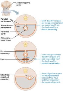
The mesentery is a double-layered peritoneum that extends from the body wall to digestive organs, providing routes for vessels and nerves, holding organs in place, and storing fat for energy and protection.
Intraperitoneal organs: Have mesentery and are located within the peritoneal cavity (e.g., stomach, most of the intestines).
Retroperitoneal organs: Lack mesentery and lie posterior to the peritoneal cavity (e.g., pancreas, parts of the duodenum).
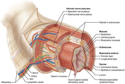
Histology of the Alimentary Canal
Layers of the Alimentary Canal
The alimentary canal is composed of four basic layers, each with distinct functions:
Mucosa: The innermost layer, a mucous membrane that lines the lumen. Functions include secretion (enzymes, mucus), absorption (nutrients), and protection (against pathogens).
Submucosa: Deep to the mucosa, this layer contains blood and lymphatic vessels, lymphoid follicles, nerve fibers, and elastic fibers for recoil after expansion.
Muscularis externa: Two layers of smooth muscle responsible for segmentation and peristalsis, aiding in movement and mixing of food.
Serosa: The outermost layer, also known as the visceral peritoneum, composed of connective tissue and epithelium.
Regulation of Digestive Activity
Control Mechanisms
Digestive activity is regulated by mechanical and chemical stimuli, nervous control, and hormonal control:
Mechanical and Chemical Stimuli: Receptors in organ walls detect stretching, solute concentration, pH, and presence of digestion end products, stimulating smooth muscle contraction and glandular activity.
Nervous Control: The enteric nervous system (a branch of the autonomic nervous system) controls digestion both intrinsically (via pacemaker cells for peristalsis and segmentation) and extrinsically (via reflex arcs to the CNS and parasympathetic output).
Hormonal Control: The stomach and small intestine produce hormones that regulate digestive processes throughout the system.
Example: When food enters the stomach, stretch receptors trigger muscle contractions and glandular secretions, while hormones like gastrin stimulate further digestive activity.
