 Back
Chapter 23: The Digestive System – Structure, Function, and Physiology
Study Guide - Smart Notes
 Tailored notes based on your materials, expanded with key definitions, examples, and context.
Overview of the Digestive System
Introduction to the Digestive System
The digestive system is responsible for breaking down food into nutrients, absorbing these nutrients into the bloodstream, and eliminating indigestible remains. It consists of the alimentary canal and accessory digestive organs, each playing a specific role in the process of digestion and absorption.
Alimentary canal (GI tract): A continuous muscular tube including the mouth, pharynx, esophagus, stomach, small intestine, and large intestine. Its main functions are digestion and absorption of food.
Accessory digestive organs: Teeth, tongue, salivary glands, liver, gallbladder, and pancreas. These organs assist in mechanical breakdown and secrete substances that aid digestion.
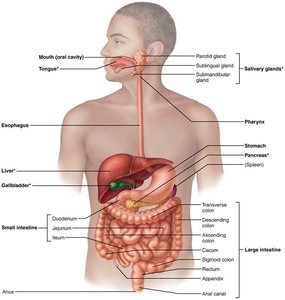
Major Processes of Digestive System Activity
Digestive system activity involves six essential processes that ensure the breakdown and absorption of nutrients:
Ingestion: Taking food into the GI tract.
Propulsion: Moving food through the GI tract, including swallowing and peristalsis (alternating waves of contraction and relaxation).
Mechanical breakdown: Chewing, mixing food with saliva, churning in the stomach, and segmentation in the intestines to increase surface area for enzymes.
Digestion: Enzymatic breakdown of complex food molecules into their chemical building blocks.
Absorption: Passage of digested fragments from the GI tract lumen into blood or lymph.
Defecation: Elimination of indigestible substances as feces.
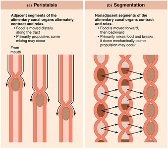
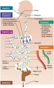
Organization and Histology of the Digestive System
Peritoneum and Mesenteries
The peritoneum is a serous membrane lining the abdominal cavity and covering abdominal organs. It consists of:
Visceral peritoneum: Covers external surfaces of digestive organs.
Parietal peritoneum: Lines the body wall.
Peritoneal cavity: Fluid-filled space between the two peritoneums, allowing organ movement and reducing friction.
Mesentery: Double layer of peritoneum that supports organs, provides routes for blood vessels, lymphatics, and nerves, and stores fat.
Retroperitoneal organs: Located posterior to the peritoneum (e.g., most of the pancreas, duodenum, parts of the large intestine).
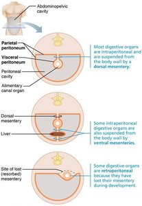
Histological Structure of the Alimentary Canal
All digestive organs share four basic layers (tunics):
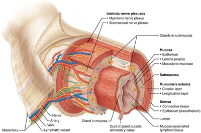
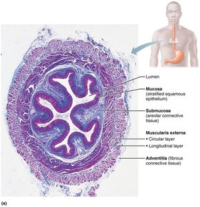
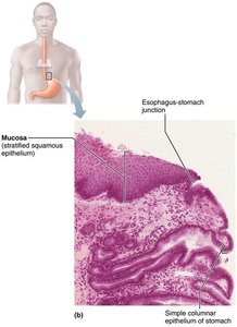
Mucosa: Innermost layer; secretes mucus, digestive enzymes, and hormones; absorbs end products; protects against infection. Composed of epithelium, lamina propria, and muscularis mucosae.
Submucosa: Areolar connective tissue with blood and lymphatic vessels, lymphoid follicles, and nerve plexuses.
Muscularis externa: Responsible for segmentation and peristalsis; consists of inner circular and outer longitudinal muscle layers. Circular layer forms sphincters in some regions.
Serosa: Outermost layer; visceral peritoneum formed from areolar connective tissue covered with mesothelium. In the esophagus, replaced by adventitia (fibrous connective tissue).
Splanchnic Circulation
Splanchnic circulation includes arteries branching from the aorta to supply digestive organs and the hepatic portal circulation, which delivers nutrient-rich blood from the GI tract to the liver for processing.
Control and Regulation of Digestive Activity
The Enteric Nervous System
The GI tract has its own nervous system, the enteric nervous system (ENS), also known as the "gut brain." It contains more neurons than the spinal cord and regulates GI tract motility and secretions.
Submucosal nerve plexus: Regulates glands and smooth muscle in the mucosa.
Myenteric nerve plexus: Controls GI tract motility.
Regulation of Digestive Activity
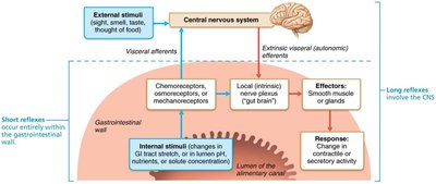
Digestive activity is regulated by mechanical and chemical stimuli, effectors (smooth muscle and glands), and control mechanisms (neurons and hormones):
Short reflexes: Mediated by the ENS, responding to stimuli within the GI tract.
Long reflexes: Involve CNS integration and autonomic nerves, responding to stimuli inside or outside the GI tract.
Parasympathetic stimulation: Enhances digestive activity.
Sympathetic stimulation: Inhibits digestive activity.
Functional Anatomy of the Digestive System
The Mouth and Associated Organs
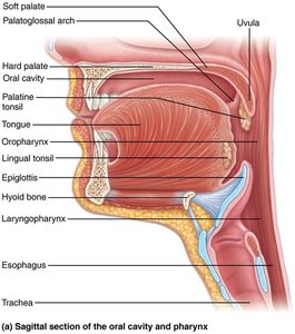
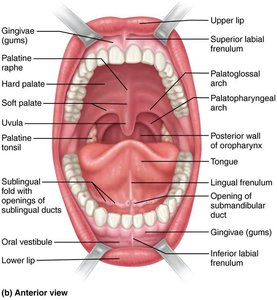
The mouth (oral cavity) is lined with stratified squamous epithelium and is bounded by the lips, cheeks, palate, and tongue. It is the site of food ingestion, mechanical breakdown, and mixing with saliva.
Lips and cheeks: Keep food between teeth during chewing.
Palate: Hard palate aids in food manipulation; soft palate closes off the nasopharynx during swallowing.
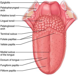
Tongue: Made of skeletal muscle; repositions food, mixes it with saliva, initiates swallowing, and aids in speech. Papillae on the tongue house taste buds.
Salivary Glands
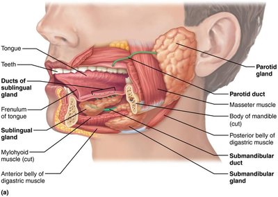
Salivary glands produce saliva, which cleanses the mouth, dissolves food chemicals, moistens food, and begins the breakdown of starch with amylase. Major glands include the parotid, submandibular, and sublingual glands.
Serous cells: Produce watery secretions with enzymes and ions.
Mucous cells: Produce mucus.
Teeth
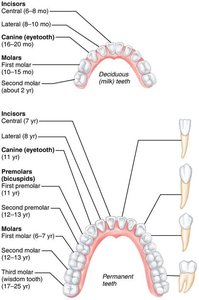
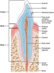
Teeth are essential for mastication (chewing), breaking food into smaller pieces. Humans have two sets of teeth: primary (deciduous) and permanent. Teeth are classified as incisors, canines, premolars, and molars, each with specific functions in cutting, tearing, and grinding food.
Pharynx and Esophagus
Structure and Function
The pharynx and esophagus serve as conduits for food and fluids from the mouth to the stomach. The pharynx is lined with stratified squamous epithelium and contains skeletal muscle for swallowing. The esophagus is a muscular tube with all four alimentary canal layers and transitions from skeletal to smooth muscle along its length.
Deglutition (Swallowing)
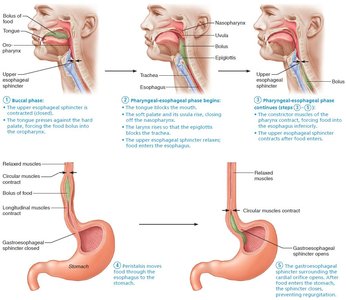
Swallowing involves two phases:
Buccal phase: Voluntary; the tongue pushes the bolus into the oropharynx.
Pharyngeal-esophageal phase: Involuntary; food is propelled through the pharynx and esophagus by coordinated muscle contractions.
Summary Table: Digestive System Overview
Process | Location | Main Function |
|---|---|---|
Ingestion | Mouth | Entry of food into the GI tract |
Propulsion | Pharynx, esophagus, stomach, intestines | Movement of food along the tract |
Mechanical breakdown | Mouth, stomach, small intestine | Chewing, churning, segmentation |
Digestion | Stomach, small intestine | Enzymatic breakdown of food |
Absorption | Small and large intestine | Transport of nutrients into blood/lymph |
Defecation | Anus | Elimination of indigestible substances |
Additional info: The digestive system is regulated by both neural and hormonal mechanisms, ensuring efficient processing and absorption of nutrients. Disorders of the digestive system can affect nutrient uptake and overall health.
