 Back
BackChapter 23: The Digestive System – Structure, Function, and Physiology
Study Guide - Smart Notes
 Tailored notes based on your materials, expanded with key definitions, examples, and context.
Overview of the Digestive System
Digestive System Functions and Organ Groups
The digestive system is responsible for breaking down food and absorbing nutrients for use by body cells. It consists of two main groups of organs:
Alimentary canal: A continuous muscular tube including the mouth, pharynx, esophagus, stomach, small intestine, and large intestine. Its primary functions are digestion and absorption of food.
Accessory digestive organs: These include the teeth, tongue, salivary glands (in the mouth), and the gallbladder, liver, and pancreas (connected by ducts). They assist in mechanical breakdown and produce secretions to aid digestion.
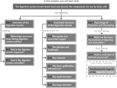
Major Processes of Digestive System Activity
Six essential activities occur during digestive system activity:
Ingestion: Taking food into the GI tract.
Propulsion: Moving food through the GI tract via swallowing and peristalsis.
Mechanical breakdown: Increasing surface area for digestion and mixing with secretions.
Digestion: Enzymatic catabolism of food into chemical components.
Absorption: Movement of digested end products from the GI lumen into blood or lymph.
Defecation: Elimination of indigestible substances via the anus.
Anatomy and Histology of the Digestive System
Peritoneum and Mesenteries
The peritoneum is a serous membrane lining the abdominopelvic cavity and covering digestive organs. It consists of:
Visceral peritoneum: Covers external surfaces of most digestive organs.
Parietal peritoneum: Lines the body wall.
Peritoneal cavity: Space between the two layers, filled with serous fluid.
Mesentery: Double layer of peritoneum that supports organs, allows passage of vessels, and stores fat.
Retroperitoneal organs: Located posterior to the mesentery, against the dorsal abdominal wall.
Layers of the Alimentary Canal
The alimentary canal has four basic layers:
Mucosa: Innermost, moist epithelial membrane; secretes, absorbs, and protects.
Submucosa: Connective tissue with blood/lymph vessels, nerve fibers, and lymphoid follicles.
Muscularis externa: Inner circular and outer longitudinal smooth muscle; responsible for peristalsis and segmentation.
Serosa: Protective outer layer (visceral peritoneum).
Splanchnic Circulation and Hepatic Portal System
Splanchnic circulation: Arteries branching from the abdominal aorta supply digestive organs.
Hepatic portal circulation: Drains nutrient-rich blood from digestive organs to the liver for processing.
Neural Control of the Digestive System
Enteric Nervous System
The GI tract has its own nervous system, the enteric nervous system, composed of enteric neurons forming two main plexuses:
Submucosal plexus: Innervates the submucosa.
Myenteric plexus: Innervates smooth muscle layers, controlling motility.
Regulation occurs via:
Short reflexes: Mediated by enteric neurons.
Long reflexes: Involve CNS integration and autonomic nerves.
Digestive activity is governed by chemical/mechanical stimuli, effectors (smooth muscle and glands), and control by neurons/hormones.
Functional Anatomy of the Digestive System
The Mouth and Associated Organs
The mouth is lined with stratified squamous epithelium and bounded by lips, cheeks, palate, and tongue. Key features:
Lips and cheeks: Skeletal muscle core, keep food between teeth.
Palate: Hard (bony) and soft (muscular); aids in food manipulation and swallowing.
Tongue: Skeletal muscle, mixes food, initiates swallowing, aids speech; papillae house taste buds.
Salivary glands: Produce saliva (water, electrolytes, enzymes, mucins, lysozyme, IgA).
Teeth: Deciduous (20) and permanent (32); classified as incisors (cutting), canines (tearing), premolars/molars (grinding).
Digestive processes in the mouth include ingestion, mechanical breakdown, propulsion, and initial digestion of polysaccharides.
Pharynx and Esophagus
The pharynx (oropharynx, laryngopharynx) and esophagus move food from the mouth to the stomach. Swallowing (deglutition) involves:
Buccal phase: Voluntary, bolus forced into oropharynx.
Pharyngeal-esophageal phase: Involuntary, food moves through pharynx and esophagus.
Stomach Structure and Function
The stomach temporarily stores food and begins protein digestion. Key features:
Regions: Cardial, fundus, body, pyloric.
Microscopic anatomy: Simple columnar epithelium, goblet cells, gastric glands (mucus, HCl, intrinsic factor, pepsinogen, hormones).
Digestive processes: Protein denaturation, absorption of alcohol/aspirin, secretion of intrinsic factor.
Regulation: Neural and hormonal mechanisms (cephalic, gastric, intestinal phases).
Alkaline tide: HCO3– pumped into blood as H+ is secreted into stomach lumen, raising blood pH.
Liver, Gallbladder, and Pancreas
Liver: Four lobes, composed of lobules (hepatocytes), secretes bile, processes nutrients, stores vitamins, detoxifies.
Gallbladder: Stores and concentrates bile.
Pancreas: Secretes pancreatic juice (enzymes and bicarbonate) into small intestine; enzymes are activated in the duodenum.
Bile and pancreatic juice delivered to intestine via ducts; secretion regulated by hormones (cholecystokinin, secretin) and neural stimuli.
Small Intestine
The small intestine is the major site for digestion and absorption. Structural modifications enhance absorption:
Divisions: Duodenum, jejunum, ileum.
Wall features: Circular folds, villi, microvilli increase surface area.
Cell types: Enterocytes (absorption), goblet cells (mucus), enteroendocrine cells (hormones), Paneth cells (antimicrobial).
Digestive processes: Segmentation, peristalsis, feedback control of gastric emptying.
Large Intestine
The large intestine absorbs water and eliminates feces. Features include:
Unique structures: Teniae coli, haustra, epiploic appendages.
Subdivisions: Cecum, appendix, colon, rectum, anal canal.
Mucosa: Thick, with mucus-producing goblet cells.
Bacteria: Ferment indigestible carbohydrates, synthesize vitamins B and K, influence immunity.
Digestive processes: Haustral contractions, mass movements, defecation reflex.
Physiology of Digestion and Absorption
Mechanism of Digestion and Absorption
Digestion hydrolyzes food into nutrients absorbed across the gut epithelium. Most digestion occurs in the small intestine via enzymatic hydrolysis. Absorption involves transport across epithelial cells into capillaries or lacteals.
Processing of Nutrients
Carbohydrates: Digested by salivary amylase, pancreatic amylase, and brush border enzymes; absorbed as monosaccharides via co-transport and facilitated diffusion.
Proteins: Digested by pepsin (stomach), pancreatic proteases, and brush border enzymes; absorbed as amino acids via co-transport and facilitated diffusion.
Lipids: Emulsified by bile salts, digested by pancreatic lipases; absorbed as monoglycerides and fatty acids, reassembled into triglycerides, packaged as chylomicrons, transported via lacteals.
Nucleic acids: Hydrolyzed by pancreatic nucleases to nucleotide monomers.
Vitamins, electrolytes, water: Small intestine absorbs dietary vitamins, electrolytes, and water (by osmosis); large intestine absorbs vitamins B and K.
Developmental Aspects of the Digestive System
Embryonic Development
Epithelial lining forms from endoderm; oral and cloacal membranes form mouth and anus.
By week 5, alimentary canal is a continuous tube.
Digestive System After Birth and Aging
Newborns: Rooting and sucking reflexes, small stomach, inefficient peristalsis.
Aging: Decline in motility, juice production, absorption; increased risk of diverticulosis, fecal incontinence, GI cancers.
Recent Discoveries and Research Topics
Mesentery classified as an organ.
Brain tanycytes control appetite.
"Good fats" may help inflammatory bowel conditions.
Gut bacteria influence peristalsis.
Suggested research topics include Helicobacter pylori infections, hepatitis, weight loss surgeries, motility disorders, digestive system cancers, and Crohn’s disease.
