 Back
BackChapter 23: The Respiratory System – Structure, Function, and Regulation
Study Guide - Smart Notes
 Tailored notes based on your materials, expanded with key definitions, examples, and context.
The Respiratory System: Overview and Organization
Introduction to the Respiratory System
The respiratory system is essential for gas exchange, supplying oxygen to body tissues and removing carbon dioxide. It supports aerobic metabolism, which is vital for cellular energy production.
Oxygen is obtained from the air via diffusion across lung surfaces.
Carbon dioxide is released from tissues and expelled through the lungs.
Blood transports these gases between the lungs and peripheral tissues.
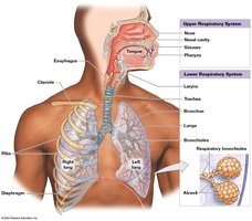
Functions of the Respiratory System
Provides a large surface area for gas exchange between air and blood.
Moves air to and from the lungs along respiratory passageways.
Protects respiratory surfaces from dehydration, temperature changes, and pathogens.
Produces sounds for communication.
Detects odors via olfactory receptors in the nasal cavity.
Organization of the Respiratory System
Anatomical divisions:
Upper respiratory system: Nose, nasal cavity, paranasal sinuses, pharynx
Lower respiratory system: Larynx, trachea, bronchi, bronchioles, alveoli
Functional divisions:
Conducting portion: Nasal cavity to larger bronchioles (air passageways)
Respiratory portion: Smallest bronchioles and alveoli (site of gas exchange)
Histology and Defense of the Respiratory Tract
Respiratory Mucosa
The respiratory mucosa lines the conducting portion and consists of:
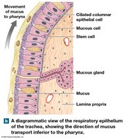
Respiratory epithelium: Innermost layer, varies by region (e.g., pseudostratified ciliated columnar in nasal cavity and trachea).
Lamina propria: Areolar tissue beneath the epithelium, contains mucous glands in upper regions and smooth muscle in lower bronchioles.
Respiratory Defense System
Mucus traps particles and pathogens.
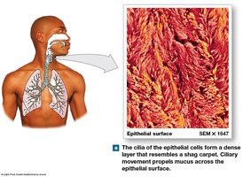
Cilia move mucus toward the pharynx (mucociliary escalator).
Alveolar macrophages engulf small particles in the alveoli.
Upper Respiratory System
Nose and Nasal Cavity
Entry point for air; nasal hairs filter large particles.
Olfactory region provides sense of smell.
Mucus moistens air; conchae create turbulence for warming, humidifying, and filtering air.
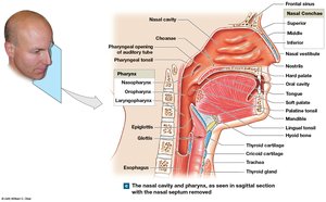
Pharynx
Shared by respiratory and digestive systems.
Divided into nasopharynx, oropharynx, and laryngopharynx.
Lined with stratified squamous epithelium for protection against abrasion.
Lower Respiratory System
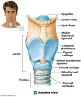
Larynx
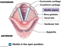
Air passes from pharynx to larynx via the glottis.
Major cartilages: thyroid, cricoid, and epiglottis.
Epiglottis prevents food/liquid from entering the airway during swallowing.
Vocal Cords
Vocal folds (true vocal cords) produce sound when air passes through the glottis.
Vestibular ligaments protect the vocal folds.
Trachea and Bronchial Tree
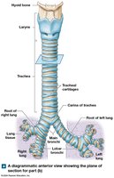
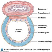
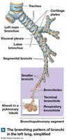
Trachea: tough, flexible tube with C-shaped cartilages to keep airway open.
Branches into right and left main bronchi, which further divide into the bronchial tree.
Bronchioles and Asthma
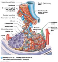
Bronchioles: smallest branches, lack cartilage, dominated by smooth muscle.
Bronchodilation (sympathetic) increases airflow; bronchoconstriction (parasympathetic or allergic reaction) decreases airflow.
Asthma: severe bronchoconstriction restricts airflow.
Gas Exchange Structures
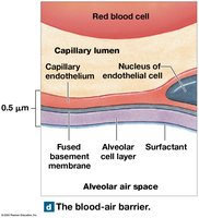
Alveoli and Blood-Air Barrier
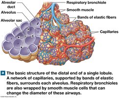
Respiratory bronchioles connect to alveoli via alveolar ducts and sacs.
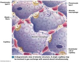
Alveoli: surrounded by elastic fibers and capillaries; site of gas exchange.
Pneumocytes type I: simple squamous cells for gas diffusion.
Pneumocytes type II: produce surfactant to reduce surface tension and keep alveoli open.
Alveolar macrophages patrol for debris and pathogens.
Pneumonia
Inflammation of lung tissue, often due to infection.
Causes swelling, fluid buildup, and impaired gas exchange.
The Lungs
Gross Anatomy of the Lungs
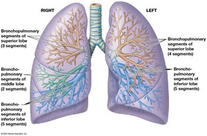
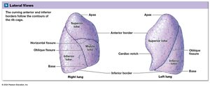
Right lung: three lobes (superior, middle, inferior).
Left lung: two lobes (superior, inferior) and a cardiac notch.
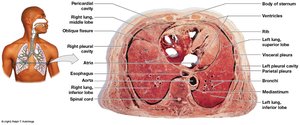
Lungs are located in pleural cavities, separated by the mediastinum.
Blood Supply and Pulmonary Embolism
Pulmonary circuit: brings deoxygenated blood to lungs for gas exchange.
Systemic circuit: supplies conducting portions of the lungs.
Pulmonary embolism: blockage of a pulmonary artery by a clot, fat, or air bubble; can cause tissue death or heart failure.
Pleural Cavities and Membranes
Pleura: serous membrane with parietal (outer) and visceral (inner) layers.
Pleural fluid lubricates and reduces friction.
Pleurisy: inflammation of pleura, causing pain due to reduced lubrication.
Respiration: External and Internal
Processes of Respiration
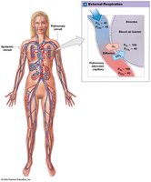
External respiration: Exchange of gases between interstitial fluid and external environment (lungs).
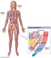
Internal respiration: Exchange of gases between blood and tissue cells.
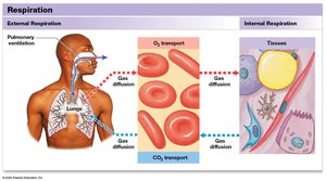
Steps in External Respiration
Pulmonary ventilation (breathing)
Gas diffusion across blood-air barrier and systemic capillaries
Transport of gases in blood
Hypoxia: Low tissue oxygen levels; Anoxia: Complete lack of oxygen, leading to cell death.
Pulmonary Ventilation
Physical Principles of Ventilation
Air moves from high to low pressure (down a pressure gradient).
Boyle’s Law: Pressure and volume of a gas are inversely related.
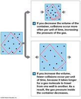
Volume Changes and Pressure Gradients
At rest: pressures inside and outside lungs are equal; no air movement.
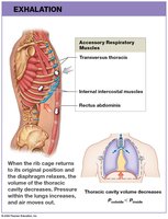
Inhalation: chest volume increases, pressure decreases, air flows in.
Exhalation: chest volume decreases, pressure increases, air flows out.
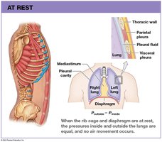
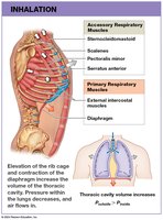
Muscles of Respiration
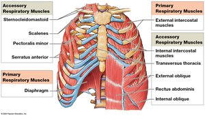
Inhalation: diaphragm contracts (flattens), external intercostals elevate ribs.
Exhalation: passive at rest (muscle relaxation); active exhalation uses internal intercostals and abdominal muscles.
Pressure Changes During Breathing
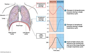
Normal atmospheric pressure: 1 atm = 760 mm Hg.
Intrapulmonary pressure: varies with inhalation/exhalation (−1 to +1 mm Hg at rest).
Intrapleural pressure: always lower than atmospheric, assists venous return to heart.
Pulmonary Volumes and Capacities
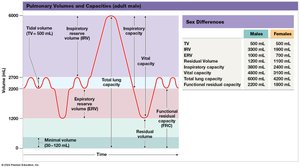
Tidal Volume (TV): Air moved per breath (average 500 mL).
Inspiratory Reserve Volume (IRV): Extra air inhaled after normal inspiration.
Expiratory Reserve Volume (ERV): Extra air exhaled after normal expiration.
Residual Volume: Air remaining after maximal exhalation.
Vital Capacity: Maximum air moved in/out in a single breath.
Total Lung Capacity: Total volume of the lungs.
Gas Exchange: Physical Principles
Partial Pressures and Gas Laws
Dalton’s Law: Each gas in a mixture exerts its own partial pressure.
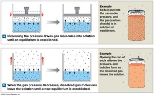
Henry’s Law: Amount of gas dissolved in a liquid is proportional to its partial pressure.
Factors Affecting Gas Exchange
Partial pressure gradients
Distance for diffusion
Lipid solubility of gases
Surface area for exchange
Ventilation-perfusion ratio
External and Internal Respiration
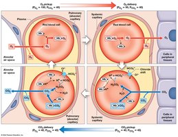
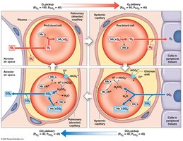
External respiration: Oxygen diffuses from alveoli to blood; carbon dioxide diffuses from blood to alveoli.
Internal respiration: Oxygen diffuses from blood to tissues; carbon dioxide diffuses from tissues to blood.
Gas Transport in Blood
Oxygen Transport
1.5% dissolved in plasma; 98.5% bound to hemoglobin (Hb).
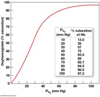
Each Hb binds up to four O2 molecules (forms oxyhemoglobin).
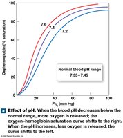
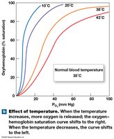
Hemoglobin saturation depends on partial pressure of O2, pH, temperature, and metabolic activity.
Carbon Dioxide Transport
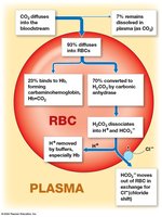
70% as bicarbonate ions (HCO3−) in plasma
23% bound to hemoglobin (carbaminohemoglobin)
7% dissolved in plasma
Control of Respiration
Local and Neural Regulation
Local factors: CO2 and O2 levels regulate blood flow and airflow in the lungs.
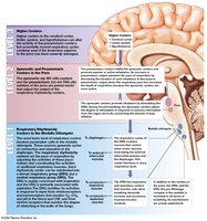
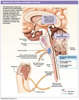
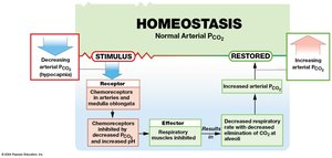
Neural control: Respiratory centers in the medulla oblongata and pons regulate rate and depth of breathing.
Voluntary control: Cerebral cortex can override involuntary centers for activities like speaking or holding breath.
Respiratory Reflexes
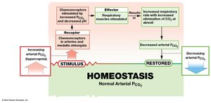
Chemoreceptors: Detect changes in CO2, O2, and pH; stimulate changes in respiratory rate.
Baroreceptors: Respond to blood pressure changes; affect respiratory rate.
Stretch receptors: Prevent overexpansion or excessive deflation of the lungs (Hering–Breuer reflexes).
Other stimuli: Pain, temperature, and visceral sensations can influence breathing.
Integration with Other Body Systems
Respiratory and Cardiovascular System Interactions
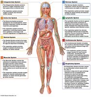
Respiratory and cardiovascular systems coordinate to maintain oxygen and carbon dioxide homeostasis.
Integration improves gas exchange efficiency, regulates blood pressure, and adjusts cardiac output as needed.
Additional info: This guide covers the structure, function, and regulation of the respiratory system, including gas exchange, transport, and integration with other organ systems, as outlined in a standard Anatomy & Physiology college course.
