 Back
BackChapter 26: The Reproductive System – Comprehensive Study Notes
Study Guide - Smart Notes
 Tailored notes based on your materials, expanded with key definitions, examples, and context.
26.1 Introduction to the Male and Female Reproductive Systems
Overview of Reproductive System Similarities
The male and female reproductive systems share several key features, including the presence of gonads (testes in males, ovaries in females), which are responsible for producing gametes and secreting sex hormones. Both systems also contain accessory reproductive organs that support the function of the gonads.
Gonads: Primary sex organs that produce gametes (sperm in males, ova in females) and secrete sex hormones (testosterone, estrogens).
Accessory Organs: Structures that aid in the transport, nourishment, and maturation of gametes.
26.1 Overview of Meiosis
Chromosome Structure and Homologous Pairs
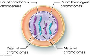
Somatic cells are diploid (2n), containing 46 chromosomes (23 pairs), with one set inherited from each parent. Each pair is termed homologous chromosomes, carrying genes for the same traits but possibly different alleles.
Alleles: Variants of a gene found at the same locus on homologous chromosomes.
Fertilization: Fusion of sperm and ovum to form a zygote with 46 chromosomes.
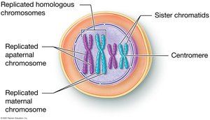
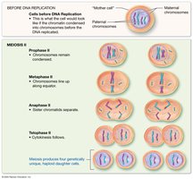
DNA Replication and Chromatin Structure
Before meiosis, DNA is replicated, resulting in chromosomes composed of two identical sister chromatids joined at a centromere. In non-dividing cells, DNA exists as chromatin.
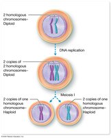
Phases of Meiosis
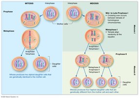
Meiosis consists of two successive divisions: Meiosis I and Meiosis II. The process reduces the chromosome number by half, producing haploid cells (1n) from diploid cells (2n).
Meiosis I: Homologous chromosomes separate, resulting in two haploid cells.
Meiosis II: Sister chromatids separate, producing four genetically unique haploid cells.
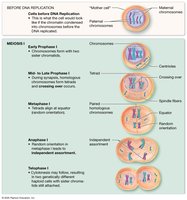
Detailed Stages of Meiosis I
Early Prophase I: Chromatin condenses, nucleoli disappear, nuclear membrane breaks down, spindle fibers form.
Mid to Late Prophase I: Homologous chromosomes pair up (synapsis) to form tetrads; crossing over occurs, exchanging genetic material and increasing genetic diversity.
Metaphase I: Tetrads align randomly at the cell equator; spindle fibers attach to centromeres.
Anaphase I: Homologous chromosomes are pulled to opposite poles (independent assortment).
Telophase I and Cytokinesis: Two haploid cells form, each with 23 chromosomes.
Detailed Stages of Meiosis II
Prophase II: Chromosomes remain condensed; nuclear envelope (if reformed) breaks down.
Metaphase II: Chromosomes align at the equator; spindle fibers attach.
Anaphase II: Sister chromatids separate and move to opposite poles.
Telophase II and Cytokinesis: Four genetically unique haploid cells are produced.
Comparison of Mitosis and Meiosis
Mitosis and meiosis are both processes of cell division, but they serve different purposes and produce different outcomes.
Mitosis: Produces two genetically identical diploid cells for growth and repair.
Meiosis: Produces four genetically unique haploid cells for sexual reproduction.
Feature | Mitosis | Meiosis |
|---|---|---|
Number of Divisions | 1 | 2 |
Number of Daughter Cells | 2 | 4 |
Genetic Identity | Identical | Unique |
Chromosome Number | Diploid (2n) | Haploid (1n) |
Function | Growth/Repair | Gamete Production |
Additional info: Crossing over and independent assortment during meiosis increase genetic diversity among offspring.
