 Back
BackChapter 27: Human Development – From Fertilization to Birth and Beyond
Study Guide - Smart Notes
 Tailored notes based on your materials, expanded with key definitions, examples, and context.
27.1 Overview of Human Development
Introduction to Human Development
Human development encompasses all events from fertilization to birth and continues through adulthood. It is divided into distinct periods based on key developmental milestones.
Pre-embryonic stage: Fertilization to implantation (first two weeks)
Embryonic stage: Conception to week 8
Fetal stage: Week 9 through birth
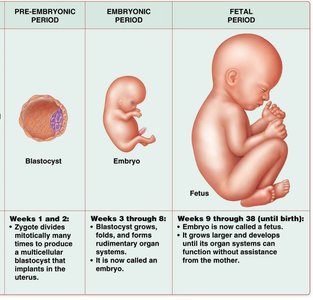
After birth, the fetus becomes a newborn, marking the beginning of the postnatal period.
27.2 Pre-embryonic Period: Fertilization through Implantation (Weeks 1 and 2)
Fertilization and Early Development
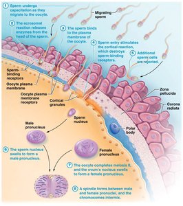
Fertilization is the process by which a sperm cell and an oocyte (egg cell) fuse to form a zygote, restoring the diploid chromosome number. This typically occurs in the uterine tube within 12–24 hours after ovulation.
Sperm capacitation: Functional maturation of sperm in the female reproductive tract.
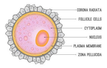
Acrosomal reaction: Release of enzymes from the sperm's acrosome to penetrate the oocyte's protective layers (corona radiata and zona pellucida).
Cortical reaction: Prevents polyspermy by hardening the zona pellucida after the first sperm enters.
Fusion of pronuclei: Male and female pronuclei merge, forming a diploid zygote.
Cleavage and Blastocyst Formation
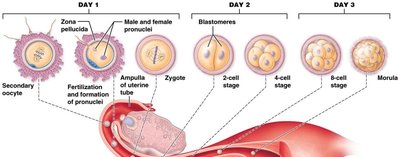
After fertilization, the zygote undergoes rapid mitotic divisions called cleavage, forming a solid ball of cells known as the morula. This structure then develops into a blastocyst, a hollow ball of cells with an outer trophoblast and an inner cell mass (embryoblast).
Morula: Solid ball of cells resulting from cleavage.
Blastocyst: Structure with an outer trophoblast (forms part of the placenta) and an inner cell mass (forms the embryo).
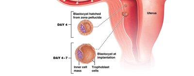
Implantation and Extraembryonic Membranes
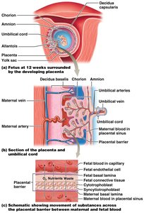
About six days after fertilization, the blastocyst implants into the endometrial lining of the uterus, which is then called the decidua. The trophoblast differentiates and contributes to the formation of the chorion, while the embryoblast forms the bilaminar disc (epiblast and hypoblast).
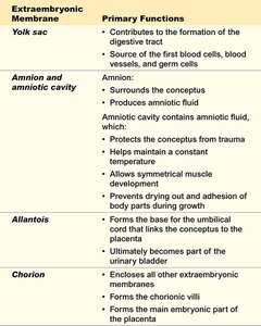
Decidua basalis: Maternal part of the placenta.
Chorion: Forms the fetal part of the placenta and secretes hCG to maintain pregnancy.
Amnion: Surrounds the embryo and contains amniotic fluid.
Yolk sac: Contributes to early blood cell formation and digestive tract development.
Allantois: Forms the base for the umbilical cord and part of the urinary bladder.
27.3 Embryonic Period: Week 3 through Week 8
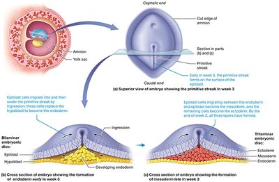
Gastrulation and Germ Layer Formation
During the third week, gastrulation transforms the bilaminar disc into a trilaminar disc, forming three primary germ layers: ectoderm, mesoderm, and endoderm. These layers give rise to all tissues and organs of the body.
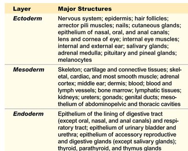
Ectoderm: Forms the nervous system, epidermis, and related structures.
Mesoderm: Forms muscle, bone, connective tissue, and the cardiovascular system.
Endoderm: Forms the lining of the digestive and respiratory tracts.
Neurulation and Organogenesis
Neurulation is the process by which the neural tube forms from the ectoderm, giving rise to the central nervous system. Organogenesis is the formation of organs from the three germ layers. By the end of the embryonic period, all major organ systems are established.
Somites: Segmented blocks of mesoderm that give rise to the vertebrae and skeletal muscles.
Placenta: Begins to develop, facilitating nutrient and waste exchange between mother and embryo.
Embryonic Growth and Differentiation (Weeks 4–8)
During weeks 4 to 8, the embryo undergoes rapid growth, folding, and differentiation. Limb buds, facial features, and organ systems become more distinct. By the end of week 8, the embryo is about 3 cm long and is now called a fetus.
27.4 Fetal Period: Week 9 until Birth (about Week 38)
Fetal Growth and Maturation
The fetal period is characterized by rapid growth and maturation of tissues and organs. Most structures are already formed; the focus is on increasing size and functional capacity.
10 weeks: Heartbeat detectable, external genitalia distinguishable.
17–20 weeks: Fetal movements felt by the mother (quickening), skin covered in vernix and lanugo.
24 weeks: Survival possible if born prematurely (with medical support).
29 weeks: Nervous system can regulate breathing and body temperature.
38 weeks: Full-term fetus, ready for birth.
27.5 Pregnancy and Childbirth
Maternal Changes During Pregnancy
Pregnancy induces significant anatomical and physiological changes in the mother, including increased uterine and fetal size, blood volume, cardiac output, and respiratory rate. Hormonal changes are crucial for maintaining pregnancy and preparing for childbirth.
hCG (human chorionic gonadotropin): Maintains corpus luteum and progesterone secretion.
Estrogen and progesterone: Maintain uterine lining and inhibit uterine contractions.
Relaxin: Softens the pubic symphysis and inhibits FSH.
hPL (human placental lactogen): Prepares mammary glands for lactation.
CRH (corticotropin-releasing hormone): Regulates timing of birth.
Labor and Delivery
Labor is initiated by hormonal changes, including increased estrogen and oxytocin levels. It consists of three stages:
Dilation: Cervix dilates to 10 cm; amniotic sac ruptures (6–12 hours).
Expulsion: Delivery of the fetus (10 minutes to 2 hours).
Placental stage: Delivery of the placenta (5–30 minutes).
27.6 Postnatal Changes in the Newborn and Mother
Newborn Adaptations
At birth, the newborn must adjust to life outside the uterus. Key changes include the initiation of breathing and closure of fetal circulatory shunts (e.g., foramen ovale).
Respiratory: First breath stimulated by increased CO2 levels.
Circulatory: Separation of pulmonary and systemic circulation.
Lactation
Milk production is stimulated by prolactin, while milk ejection (let-down reflex) is triggered by oxytocin in response to suckling. Progesterone inhibits prolactin during pregnancy, but after birth, prolactin levels rise to support lactation.
Summary Table: Major Events of Human Development
Period | Major Events |
|---|---|
Pre-embryonic (Weeks 1–2) | Fertilization, cleavage, blastocyst formation, implantation |
Embryonic (Weeks 3–8) | Gastrulation, neurulation, organogenesis, placenta formation |
Fetal (Week 9–Birth) | Growth, tissue and organ maturation, preparation for birth |
