 Back
BackChapter 3: Cells – The Living Units (ANP College Study Guide)
Study Guide - Smart Notes
 Tailored notes based on your materials, expanded with key definitions, examples, and context.
Cells: The Living Units
Introduction to Cells
Cells are the fundamental structural and functional units of life. Every organism is composed of cells, which carry out essential biological processes. The diversity in cell structure reflects the diversity in cell function, with over 250 distinct types of human cells.
Definition: A cell is the smallest unit capable of performing all vital physiological functions.
Cell Diversity: Cells vary in shape, size, and function, such as epithelial cells (lining), erythrocytes (transport), muscle cells (movement), fat cells (storage), macrophages (defense), nerve cells (information processing), and sperm cells (reproduction).
Example: Erythrocytes transport oxygen, while nerve cells transmit electrical signals.
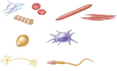
Additional info: Cell specialization is essential for multicellular organisms, allowing division of labor and complex body functions.
Structure of the Generalized Cell
Main Regions of a Cell
A typical cell consists of three major regions: the plasma membrane, cytoplasm, and nucleus. Each region has distinct structural and functional roles.
Plasma Membrane: Encloses the cell, regulates entry and exit of substances, and facilitates communication.
Cytoplasm: Contains cytosol, organelles, and inclusions; site of metabolic activities.
Nucleus: Houses genetic material (DNA), controls cellular activities, and is involved in cell division.
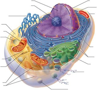
Additional info: The cytoplasm includes specialized organelles such as mitochondria (energy production), ribosomes (protein synthesis), endoplasmic reticulum (protein and lipid synthesis), Golgi apparatus (modification and packaging), lysosomes (digestion), and peroxisomes (detoxification).
Plasma Membrane: Structure and Function
Functions of the Plasma Membrane
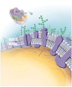
The plasma membrane is a dynamic boundary that separates the cell's internal environment from the external environment. Its structure enables selective permeability, communication, and cell recognition.
Physical Barrier: Maintains cellular integrity by enclosing cytoplasm.
Selective Permeability: Controls which substances can enter or exit the cell.
Communication: Membrane proteins relay signals from external messengers to the cell interior.
Cell Recognition: Glycocalyx (carbohydrate layer) enables cells to identify each other.
Chemical Composition of the Plasma Membrane
The plasma membrane is primarily composed of lipids, proteins, and carbohydrates. The arrangement and properties of these molecules determine membrane function.
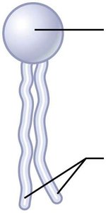
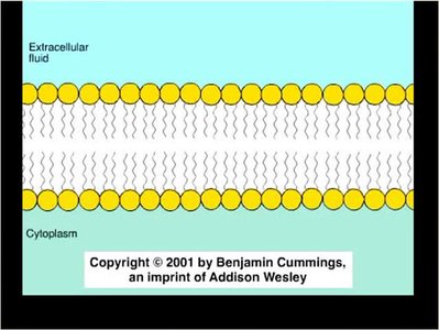
Phospholipids: Form a bilayer with hydrophilic heads facing outward and hydrophobic tails inward, creating a barrier to water-soluble substances.
Cholesterol: Stabilizes the membrane and modulates fluidity.
Proteins: Integral and peripheral proteins serve as transporters, receptors, and anchors.
Carbohydrates: Attached to proteins (glycoproteins) or lipids (glycolipids), forming the glycocalyx for cell recognition.
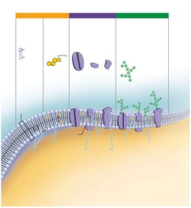
Additional info: The fluid mosaic model describes the dynamic nature of the membrane, with lipids and proteins moving laterally within the bilayer.
Lipid Bilayer Structure
The phospholipid bilayer is the fundamental structure of the plasma membrane, providing both flexibility and selective permeability.
Hydrophilic Heads: Face the aqueous environments inside and outside the cell.
Hydrophobic Tails: Face inward, away from water, forming a barrier to polar molecules.
Membrane Fluidity and Selective Permeability
The plasma membrane is fluid, allowing movement of lipids and proteins. Selective permeability is achieved through the specific arrangement of membrane components.
Fluid Mosaic Model: Membrane components move within the bilayer, enabling flexibility and self-healing.
Cholesterol: Reduces fluidity and increases stability.
Cell Junctions
Tight Junctions
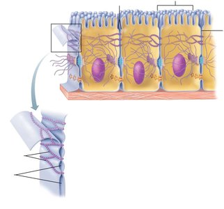
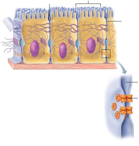
Tight junctions are specialized connections between adjacent cells that create a seal, preventing the passage of molecules between cells.
Structure: Interlocking junctional proteins form continuous seals.
Function: Impermeable barrier; essential in epithelial tissues.
Desmosomes
Desmosomes are anchoring junctions that bind cells together, providing mechanical strength and preventing tearing.
Structure: Linker proteins (cadherins) and intermediate filaments (keratin) connect cells.
Function: Mechanical stability; found in tissues subject to stress (e.g., skin, heart).
Gap Junctions
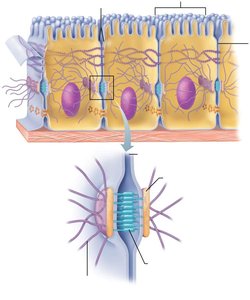
Gap junctions are communicating junctions that allow ions and small molecules to pass directly between cells, facilitating rapid communication.
Structure: Connexons form channels between adjacent cells.
Function: Electrical and chemical communication; important in cardiac and embryonic cells.
Gradients Across the Plasma Membrane
Types of Gradients
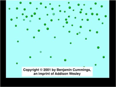
Gradients are differences in concentration or charge across the plasma membrane, driving the movement of molecules.
Concentration Gradient: Difference in chemical concentration across the membrane.
Electrical Gradient: Difference in ion concentration, creating a voltage.
Electrochemical Gradient: Combined effect of concentration and electrical gradients.
Additional info: Gradients are essential for processes such as nerve impulse transmission and muscle contraction.
Transport Across the Plasma Membrane
Passive Transport
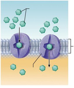
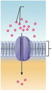
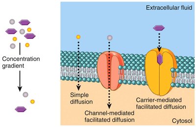
Passive transport involves the movement of substances across the membrane without energy input, relying on concentration gradients.
Simple Diffusion: Movement of small, nonpolar molecules directly through the lipid bilayer.
Facilitated Diffusion: Movement of larger or polar molecules via protein channels or carriers.
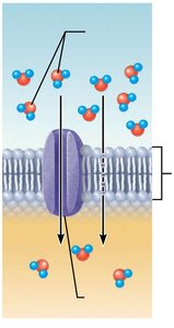
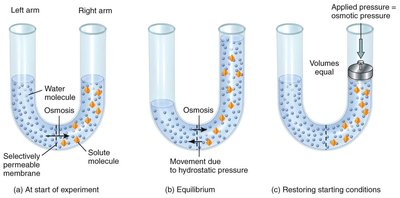
Osmosis: Diffusion of water through the membrane or aquaporins.
Osmosis and Osmotic Pressure
Osmosis is the movement of water across a selectively permeable membrane. Osmotic pressure is the force required to prevent water movement.
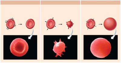
Isotonic Solution: Equal solute concentration; cells retain normal shape.
Hypertonic Solution: Higher solute concentration outside; cells lose water and shrink.
Hypotonic Solution: Lower solute concentration outside; cells gain water and may burst.
Active Transport
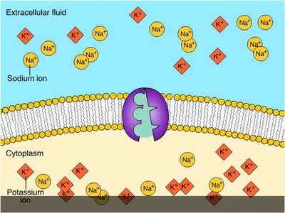
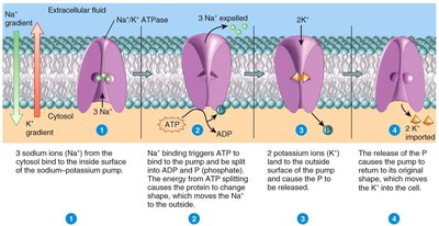
Active transport requires energy (ATP) to move substances against their concentration gradients. It is essential for maintaining cellular homeostasis.
Primary Active Transport: Direct use of ATP to transport molecules (e.g., sodium-potassium pump).
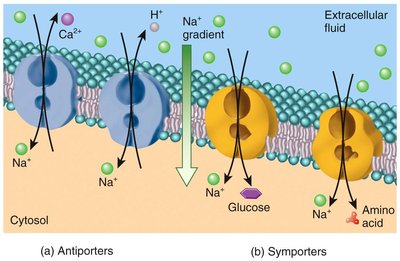
Secondary Active Transport: Uses energy stored in gradients to transport other substances.
Vesicular Transport
Vesicular transport involves the movement of large particles, macromolecules, and fluids across the membrane via vesicles.
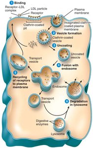
Endocytosis: Uptake of substances into the cell. Includes pinocytosis (fluid uptake), phagocytosis (particle uptake), and receptor-mediated endocytosis (specific uptake).
Exocytosis: Release of substances from the cell by fusion of vesicles with the plasma membrane.
Transcytosis: Combination of endocytosis and exocytosis to move substances across the cell.
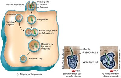
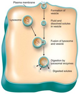
Additional info: Vesicular transport is crucial for immune responses, neurotransmitter release, and nutrient absorption.
Summary Table: Types of Membrane Transport
Transport Type | Energy Required | Direction | Substances Transported | Mechanism |
|---|---|---|---|---|
Simple Diffusion | No | High to Low | Small, nonpolar molecules | Direct through lipid bilayer |
Facilitated Diffusion | No | High to Low | Polar molecules, ions | Via protein channels/carriers |
Osmosis | No | High to Low (water) | Water | Through bilayer or aquaporins |
Primary Active Transport | Yes (ATP) | Low to High | Ions (e.g., Na+, K+) | Pump proteins |
Secondary Active Transport | Indirect (gradient) | Low to High | Glucose, amino acids | Symporters/antiporters |
Endocytosis | Yes | Into cell | Large particles, fluids | Vesicle formation |
Exocytosis | Yes | Out of cell | Macromolecules | Vesicle fusion |
Key Equations
Diffusion Rate:
Osmotic Pressure:
Sodium-Potassium Pump: per ATP hydrolyzed
Conclusion
Understanding cell structure and membrane transport is fundamental to the study of anatomy and physiology. The plasma membrane's selective permeability, cell junctions, and transport mechanisms ensure cellular function and homeostasis.
