 Back
BackChapter 3: Cells – The Living Units (BSC 2085 Human Anatomy and Physiology I)
Study Guide - Smart Notes
 Tailored notes based on your materials, expanded with key definitions, examples, and context.
Cells: The Living Units
Overview of Cells
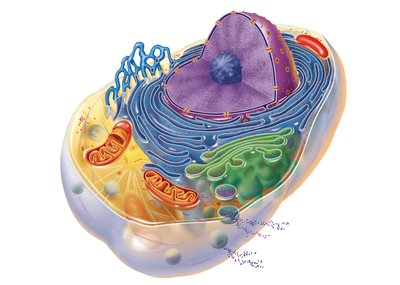
Cells are the basic structural and functional units of all living organisms. Human cells share common structures but can be highly specialized for different functions. Each cell consists of three main parts: the plasma membrane, cytoplasm, and nucleus.
Plasma membrane: Flexible outer boundary that separates the cell from its environment.
Cytoplasm: Intracellular fluid containing organelles.
Nucleus: Control center containing genetic material.
The Plasma Membrane
Structure and Function
The plasma membrane is a dynamic, bimolecular layer of lipids and proteins, often described as a fluid mosaic. It separates intracellular fluid (ICF) from extracellular fluid (ECF), including interstitial fluid (IF) that surrounds cells. The membrane is selectively permeable, allowing some substances to pass while restricting others.
Phospholipid bilayer: Main structural component, with hydrophilic (polar) heads facing outward and hydrophobic (nonpolar) tails facing inward.
Cholesterol: Stabilizes membrane fluidity.
Glycolipids: Lipids with attached sugar groups, found on the outer surface.
Membrane Proteins
Membrane proteins are essential for various cellular functions and are classified as integral or peripheral proteins.
Integral proteins: Embedded within the lipid bilayer; function as transporters, receptors, or enzymes.
Peripheral proteins: Loosely attached to membrane surfaces; involved in support, cell signaling, and forming part of the glycocalyx.
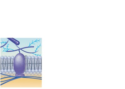
Functions of Membrane Proteins
Transport: Channels and carriers move substances across the membrane.
Receptors for signal transduction: Bind signaling molecules and initiate cellular responses.
Attachment to cytoskeleton and ECM: Maintain cell shape and stabilize membrane location.
Enzymatic activity: Catalyze metabolic reactions.
Intercellular joining: Form cell junctions for communication and adhesion.
Cell-cell recognition: Glycoproteins serve as identification tags.
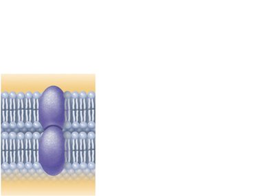
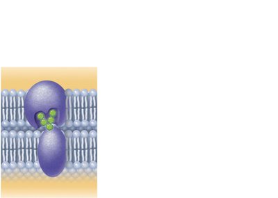
Membrane Junctions
Cells are connected by specialized junctions that regulate communication and adhesion:
Tight junctions: Prevent passage of molecules between cells.
Desmosomes: Anchor cells together, providing mechanical stability.
Gap junctions: Allow ions and small molecules to pass directly between cells, facilitating communication.
Membrane Transport
Types of Membrane Transport
Transport across the plasma membrane is classified as passive (no energy required) or active (requires ATP).
Passive processes: Substances move down their concentration gradient (e.g., diffusion, osmosis).
Active processes: Substances move against their concentration gradient using energy (e.g., active transport, vesicular transport).
Passive Transport
Simple diffusion: Movement of lipid-soluble molecules directly through the phospholipid bilayer.
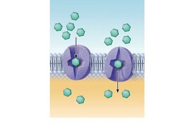
Facilitated diffusion: Movement of lipid-insoluble molecules via protein carriers or channels.
Osmosis: Diffusion of water through a selectively permeable membrane.
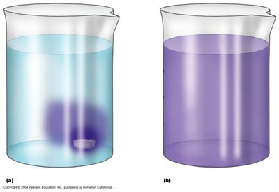
Osmosis and Tonicity
Osmosis is the movement of water across a membrane toward higher solute concentration. Tonicity describes how a solution affects cell volume:
Isotonic: No net water movement; cell volume remains constant.
Hypertonic: Water leaves the cell; cell shrinks.
Hypotonic: Water enters the cell; cell swells and may burst.
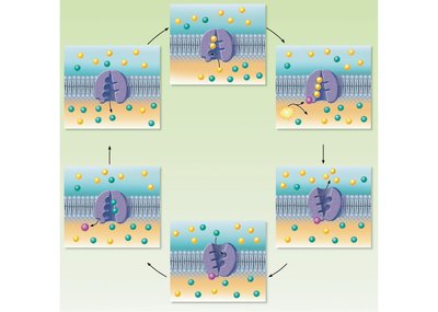
Active Transport
Active transport uses carrier proteins (pumps) and ATP to move substances against their concentration gradients. The sodium-potassium pump (Na+/K+ ATPase) is a key example, maintaining cellular ion balance.
Vesicular Transport
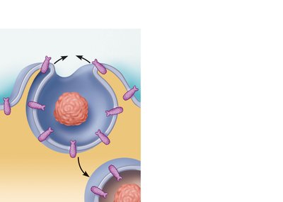
Vesicular transport moves large particles and macromolecules across membranes via vesicles. Types include:
Endocytosis: Uptake of materials into the cell (e.g., phagocytosis, pinocytosis).
Exocytosis: Release of materials from the cell.
Cytoplasm and Organelles
Cytoplasm
The cytoplasm is the cellular material between the plasma membrane and nucleus. It consists of cytosol (fluid), organelles (metabolic machinery), and inclusions (stored nutrients or pigments).
Major Organelles
Mitochondria: Site of ATP production via aerobic respiration; contain their own DNA and RNA.
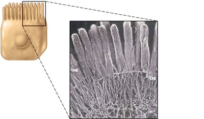
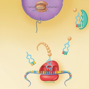
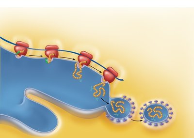
Ribosomes: Sites of protein synthesis; free ribosomes produce cytosolic proteins, while membrane-bound ribosomes synthesize proteins for membranes or export.
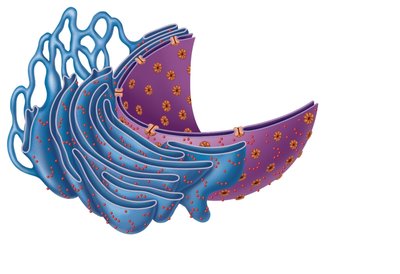
Endoplasmic Reticulum (ER): Network of membranes; rough ER (with ribosomes) synthesizes proteins, smooth ER is involved in lipid metabolism and detoxification.
Golgi Apparatus: Modifies, sorts, and packages proteins and lipids for secretion or delivery to other organelles.
Lysosomes: Contain digestive enzymes to break down waste and cellular debris.
Peroxisomes: Detoxify harmful substances and neutralize free radicals.
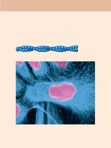
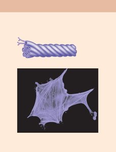
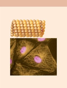
Cytoskeleton: Network of protein filaments (microfilaments, intermediate filaments, microtubules) providing structural support and facilitating movement.
Centrosome and Centrioles: Organize microtubules and are essential for cell division.
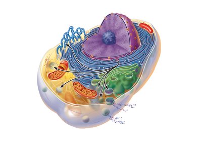
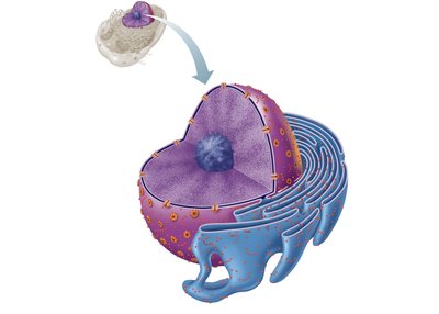
The Nucleus
Structure and Function
The nucleus is the cell's control center, containing DNA and directing cellular activities. It is surrounded by a double-membrane nuclear envelope with pores for molecular transport. The nucleolus within the nucleus is the site of ribosomal RNA synthesis and ribosome assembly.
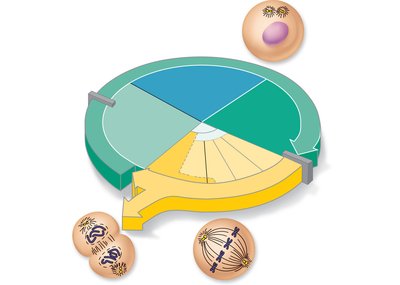
The Cell Life Cycle (Mitosis)
Phases of the Cell Cycle
The cell cycle consists of interphase (growth and DNA replication) and the mitotic phase (cell division). Interphase includes G1 (growth), S (DNA synthesis), and G2 (preparation for division).

Mitosis
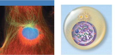
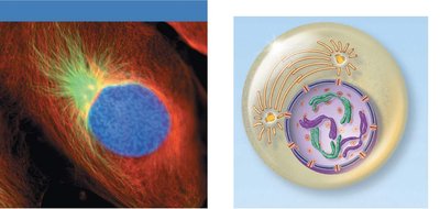
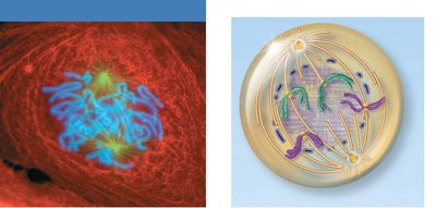
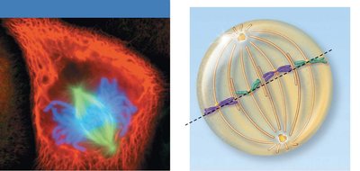
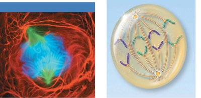
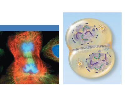
Mitosis is the process of nuclear division, producing two identical daughter cells. It consists of four stages:
Prophase: Chromosomes condense, spindle fibers form, nuclear envelope breaks down.
Metaphase: Chromosomes align at the metaphase plate.
Anaphase: Sister chromatids separate and move to opposite poles.
Telophase: Chromosomes decondense, nuclear envelopes reform.
Cytokinesis (division of the cytoplasm) overlaps with telophase, resulting in two separate cells.
DNA Structure and Function
DNA Organization
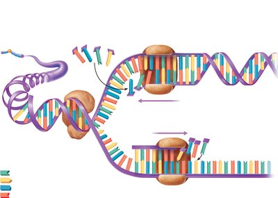
DNA is a double helix composed of nucleotides (deoxyribose sugar, phosphate group, nitrogenous base). The sequence of bases encodes genetic information. DNA is packaged into chromatin and chromosomes within the nucleus.
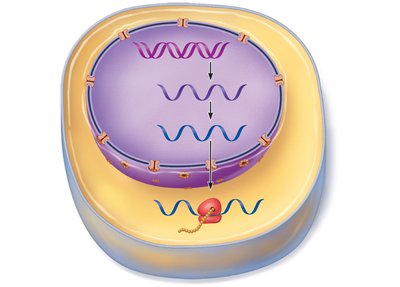
Gene Expression: Transcription and Translation
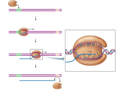
Transcription: Synthesis of mRNA from a DNA template in the nucleus.
Translation: Synthesis of proteins from mRNA at the ribosome in the cytoplasm, involving tRNA and rRNA.
Additional info: This guide covers the essential concepts of cell structure, membrane dynamics, organelle function, and the cell cycle, as outlined in a typical introductory anatomy and physiology course. The included images reinforce key structural and functional aspects of cells and their components.
