 Back
BackChapter 3: The Cell – Structure, Function, and Components
Study Guide - Smart Notes
 Tailored notes based on your materials, expanded with key definitions, examples, and context.
Chapter 3: The Cell
Introduction to the Cell
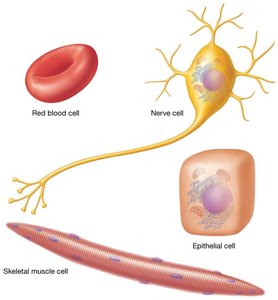
The cell is the fundamental unit of life in all organisms. Cells are separated from their environment by a plasma membrane and vary in shape, size, content, and function. Understanding the structure and function of cells is essential for comprehending the organization and physiology of the human body.
Definition: The cell is the smallest structural and functional unit of an organism.
Key Functions: Metabolism, substance transport, communication, and reproduction.
Types of Cells: Prokaryotic (no nucleus, e.g., bacteria) and Eukaryotic (with nucleus, e.g., animal cells).
Planes and Directions in Anatomy
Understanding anatomical planes and directional terms is crucial for describing locations and relationships within the body.
Sagittal Plane: Divides the body into right and left portions.
Frontal (Coronal) Plane: Divides the body into anterior and posterior portions.
Transverse Plane: Divides the body into superior and inferior portions.
Oblique Plane: Passes through the body at an angle.
Directional Terms: Superior (above), Inferior (below), Anterior (front), Posterior (back), Lateral (away from midline), Medial (toward midline).
Example Application: The liver is superior to the stomach.
Cell Structure and Components
Basic Components of Animal Cells
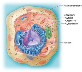
Animal cells have three primary components: the plasma membrane, cytoplasm, and nucleus. Each component plays a specific role in maintaining cellular function and integrity.
Plasma Membrane: Separates the cell from its environment and regulates transport.
Cytoplasm: Contains cytosol, organelles, and the cytoskeleton.
Nucleus: Directs cellular activities and contains genetic material.
Plasma Membrane: Structure and Function
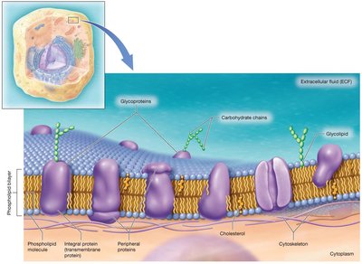
The plasma membrane, also known as the cell membrane, is a dynamic structure that isolates the cell, provides support, communicates with other cells, and regulates the movement of substances.
Structure: Composed of a phospholipid bilayer with embedded proteins, cholesterol, and carbohydrates.
Fluid Mosaic Model: Describes the membrane as a flexible layer with various proteins and molecules floating within it.
Functions: Physical isolation, structural support, communication, selective transport, and cell identification.
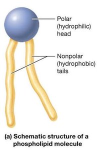
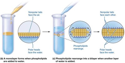
Phospholipid Bilayer
The phospholipid bilayer forms the main framework of the cell membrane, creating a barrier between the extracellular fluid (ECF) and the cytosol.
Hydrophilic Heads: Face outward toward water.
Hydrophobic Tails: Face inward, away from water.
Self-Assembly: In aqueous environments, phospholipids arrange into a bilayer to protect hydrophobic tails from water.
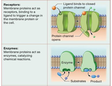
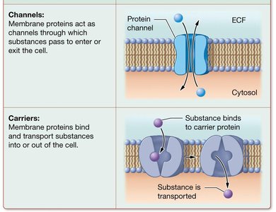
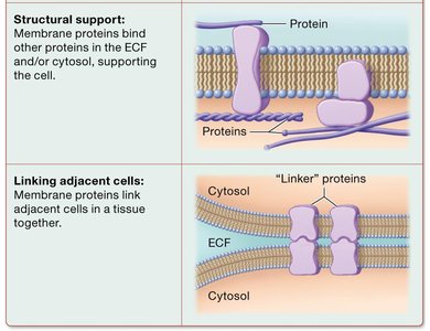
Membrane Proteins
Membrane proteins are essential for the diverse functions of the plasma membrane. They are classified as integral (span the membrane) or peripheral (attached to the surface).
Integral Proteins: Involved in transport, signaling, and adhesion.
Peripheral Proteins: Involved in signaling, structural support, and maintaining cell shape.
Functional Types: Channels, carriers, receptors, enzymes, structural support proteins, and linker proteins.
Cholesterol and Carbohydrates
Cholesterol stabilizes the plasma membrane, maintaining its fluidity and flexibility. Carbohydrates, present as glycolipids and glycoproteins, are involved in cell recognition and communication.
Cholesterol: Prevents the membrane from becoming too rigid or too fluid with temperature changes.
Carbohydrates: Aid in cell recognition and signaling.
Membrane Junctions
Membrane junctions connect cells, seal boundaries, and facilitate communication.
Tight Junctions: Form impermeable barriers (e.g., in epithelial cells).
Gap Junctions: Allow passage of ions and small molecules for communication (e.g., in cardiac muscle).
Anchoring Junctions (Desmosomes): Provide mechanical stability (e.g., in skin and intestines).
Cytoplasm and Its Components
Cytoplasm
The cytoplasm is the cellular material inside the plasma membrane and outside the nucleus. It is the site of most cellular activities and consists of cytosol, organelles, and inclusions.
Cytosol: The fluid portion containing water, proteins, salts, and sugars.
Organelles: Specialized structures performing specific functions.
Inclusions: Stored nutrients and pigments.
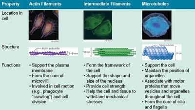
Cytoskeleton
The cytoskeleton is a network of protein filaments that provides structural support, maintains cell shape, and facilitates movement.
Actin Filaments (Microfilaments): Support the plasma membrane, form the core of microvilli, and are involved in cell movement and division.
Intermediate Filaments: Form the framework of the cell, support the nucleus, and provide cell strength.
Microtubules: Maintain organelle position, form the core of cilia and flagella, and are involved in intracellular transport.

Organelles: Structure and Function
Membrane-Bound Organelles
Mitochondrion: Double-membraned organelle responsible for ATP synthesis via oxidative catabolism. Contains its own DNA and ribosomes.
Peroxisome: Membrane-enclosed organelle that detoxifies chemicals, metabolizes fatty acids, and synthesizes certain phospholipids.
Endoplasmic Reticulum (ER): Network of membranes; Rough ER (RER) is studded with ribosomes and synthesizes proteins, while Smooth ER (SER) synthesizes lipids, stores calcium, and detoxifies substances.
Golgi Apparatus: Stack of flattened sacs that sorts, modifies, and packages proteins and lipids for transport.
Lysosome: Membrane-enclosed organelle containing digestive enzymes for breaking down waste and cellular debris.
Non-Membrane-Bound Organelles
Ribosome: Site of protein synthesis; composed of rRNA and proteins. Can be free in the cytosol or bound to the ER.
Centrosome: Organizes microtubules and is important for cell division.
Nucleus: Structure and Function
Nucleus
The nucleus is the control center of the cell, containing DNA, RNA, and nuclear proteins. It is separated from the cytoplasm by a double-membraned nuclear envelope.
Nuclear Envelope: Double lipid bilayer with nuclear pores for transport.
Nucleolus: Site of rRNA synthesis and ribosome assembly.
Chromatin: DNA-protein complex that condenses into chromosomes during cell division.
Chromosomes: Highly condensed chromatin visible during cell division; humans have 46 chromosomes (23 pairs).
Summary Table: Major Cell Structures and Functions
Structure | Main Function |
|---|---|
Plasma Membrane | Selective barrier, communication, support |
Cytoplasm | Site of metabolic activity |
Cytoskeleton | Shape, support, movement |
Mitochondrion | ATP synthesis |
Ribosome | Protein synthesis |
ER (Rough/Smooth) | Protein/lipid synthesis, detoxification |
Golgi Apparatus | Protein modification and packaging |
Lysosome | Digestion and recycling |
Nucleus | Genetic control, RNA synthesis |
