 Back
Chapter 3: The Cell – Structure, Function, and Division
Study Guide - Smart Notes
 Tailored notes based on your materials, expanded with key definitions, examples, and context.
Chap 3 part II - The Cell - Chapter 3 Part II
Introduction to the Cell
The cell is the basic structural and functional unit of all living organisms. Cells contain specialized structures called organelles, each with distinct roles that contribute to cellular and organismal homeostasis.
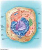
Cytoplasmic Organelles
Membrane-Enclosed vs. Non-Membrane-Enclosed Organelles
Membrane-Enclosed Organelles: Surrounded by a phospholipid bilayer; includes mitochondria, peroxisomes, endoplasmic reticulum (ER), Golgi apparatus, and lysosomes.
Non-Membrane-Enclosed Organelles: Not surrounded by a membrane; includes ribosomes and centrosomes.
Mitochondria
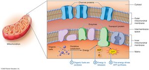
Mitochondria are the cell’s primary site for ATP production through oxidative catabolism. Cells with high energy demands contain more mitochondria. Each mitochondrion has its own DNA, enzymes, and ribosomes, and is composed of a smooth outer membrane and a highly folded inner membrane (cristae).
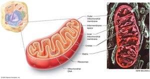
The outer membrane contains large channels for molecule passage, while the inner membrane is selective and contains transport proteins. The matrix, the innermost compartment, houses mitochondrial DNA and enzymes for ATP production.
Peroxisomes
Use oxygen to oxidize and strip hydrogen from organic compounds, producing hydrogen peroxide (H2O2).
Detoxify harmful substances (e.g., alcohol), break down fatty acids, and synthesize certain phospholipids.
Ribosomes
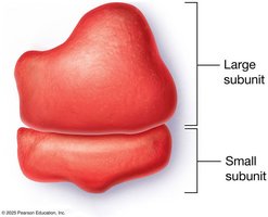
Ribosomes are the sites of protein synthesis, composed of ribosomal RNA (rRNA) and proteins. They exist as free ribosomes in the cytosol or bound to membranes (e.g., rough ER).
The Endomembrane System
Overview
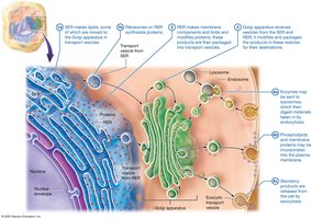
The endomembrane system is a network of organelles that synthesize, modify, and transport cellular products via vesicles. It includes the plasma membrane, endoplasmic reticulum (ER), Golgi apparatus, lysosomes, and the nuclear envelope.
Endoplasmic Reticulum (ER)
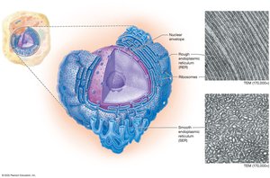
Rough ER (RER): Studded with ribosomes; folds polypeptides, recognizes misfolded proteins, and synthesizes membrane and secretory proteins.
Smooth ER (SER): Lacks ribosomes; stores calcium ions, detoxifies chemicals, and synthesizes lipids and steroid hormones.
Golgi Apparatus
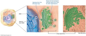
The Golgi apparatus consists of flattened sacs that modify, sort, and package proteins and lipids from the ER for secretion or delivery to other organelles.
Lysosomes
Lysosomes are acidic, membrane-bound sacs containing acid hydrolases that digest macromolecules, old organelles, and pathogens. They play a key role in autophagy and immune defense.
Summary of Endomembrane System Function
Cytoskeleton
Structure and Function
The cytoskeleton is a network of protein filaments that provides structural support, maintains cell shape, anchors organelles, and enables movement. It consists of three main types of filaments:
Actin Filaments (Microfilaments): Thin, flexible filaments involved in cell movement and shape maintenance.
Intermediate Filaments: Rope-like, provide mechanical strength and stabilize cell structure.
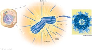
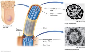
Microtubules: Hollow tubes that maintain cell architecture, serve as tracks for organelle movement, and form the core of cilia and flagella.
Centrosome and Centrioles
The centrosome is the microtubule-organizing center, containing a pair of centrioles that play a role in cell division.
Cellular Extensions
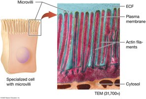
Microvilli: Actin-supported projections that increase surface area for absorption (e.g., in the small intestine).
Cilia and Flagella: Microtubule-based structures for movement; cilia move substances across cell surfaces, while flagella propel entire cells (e.g., sperm).
The Nucleus
Structure and Function
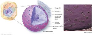
The nucleus directs cellular activities and houses DNA. It consists of the nuclear envelope, nucleoplasm, and nucleolus (site of ribosome assembly).
Nuclear Envelope and Pores
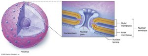
The nuclear envelope is a double membrane with nuclear pores that regulate molecular traffic between the nucleus and cytoplasm.
Chromatin and Chromosomes
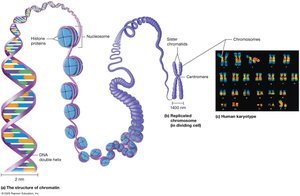
DNA is packaged with histone proteins into chromatin. During cell division, chromatin condenses into chromosomes, each consisting of two sister chromatids joined at a centromere. Humans have 46 chromosomes (23 pairs).
Protein Synthesis
Overview
Protein synthesis is the process by which cells build proteins based on genetic instructions. It involves two main steps: transcription and translation.

The Genetic Code
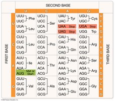
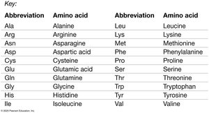
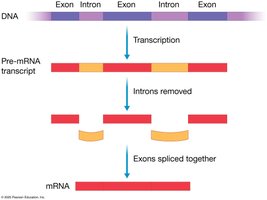
Genes are DNA segments that code for proteins. The genetic code is a set of rules by which nucleotide triplets (codons) in mRNA specify amino acids. Exons are coding regions; introns are noncoding. Mutations are changes in DNA sequence that can lead to disease.
Transcription
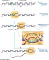
Transcription is the synthesis of mRNA from a DNA template. It occurs in three stages: initiation, elongation, and termination. The resulting pre-mRNA is processed by removing introns and splicing exons together.
Translation
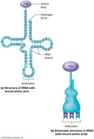
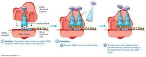
Translation is the process by which ribosomes synthesize a polypeptide using the mRNA sequence. tRNA molecules bring specific amino acids to the ribosome, matching their anticodon to the mRNA codon. Translation proceeds through initiation, elongation, and termination.
The Cell Cycle
Phases of the Cell Cycle
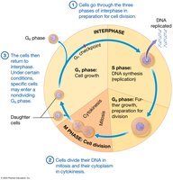
The cell cycle is the series of events from cell formation to division. It consists of interphase (G1, S, G2) and the M phase (mitosis and cytokinesis).
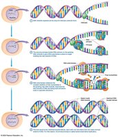
DNA Replication
During S phase, DNA is replicated by semiconservative replication, ensuring each daughter cell receives an identical genome. Key enzymes include helicase, primase, and DNA polymerase.
Mitosis and Cytokinesis
Mitosis divides the genetic material into two nuclei, while cytokinesis splits the cytoplasm, producing two identical daughter cells. The stages of mitosis are prophase, metaphase, anaphase, and telophase.
Cell Cycle Control and Cancer
Cell division is tightly regulated by checkpoints and extracellular signals. Failure to regulate the cell cycle can result in uncontrolled cell growth (tumors). Benign tumors remain localized, while malignant (cancerous) tumors can invade other tissues (metastasis).
Summary Table: Cytoplasmic Organelles
Organelle | Structure | Function |
|---|---|---|
Mitochondrion | Double membrane, cristae, matrix | ATP production via oxidative catabolism |
Peroxisome | Membrane-bound sac | Oxidizes toxins, breaks down fatty acids, synthesizes phospholipids |
Ribosome | Two subunits (rRNA + protein) | Protein synthesis |
Rough ER | Membranous, ribosome-studded | Protein folding, modification, and transport |
Smooth ER | Membranous, no ribosomes | Lipid synthesis, detoxification, calcium storage |
Golgi Apparatus | Stack of flattened sacs | Modifies, sorts, and packages proteins/lipids |
Lysosome | Membrane-bound sac with enzymes | Digestion of macromolecules, autophagy |
