 Back
Chapter 3: Tissues – Structure, Function, and Classification
Study Guide - Smart Notes
 Tailored notes based on your materials, expanded with key definitions, examples, and context.
Introduction to Tissues (Histology)
Overview of Tissues
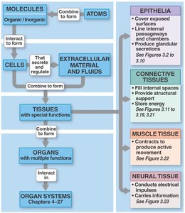
Histology is the study of tissues, which are collections of specialized cells and cell products organized to perform specific functions. The human body is composed of four primary tissue types: epithelial, connective, muscle, and nervous tissue. Each type has distinct structural and functional characteristics essential for maintaining homeostasis and supporting life.
Epithelial tissue: Covers surfaces, lines cavities, and forms glands.
Connective tissue: Provides support, binds tissues, stores energy, and transports substances.
Muscle tissue: Responsible for movement through contraction.
Nervous tissue: Conducts electrical impulses and processes information.
Epithelial Tissue
General Characteristics
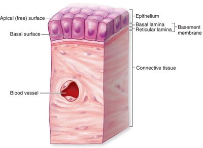
Epithelial tissue forms the linings of internal and external body surfaces and constitutes the functional tissue of glands. It is characterized by closely packed cells with minimal extracellular matrix, polarity (distinct apical and basal surfaces), avascularity (lacking blood vessels), and high regenerative capacity.
Polarity: Apical (free) surface faces the lumen or exterior; basal surface attaches to underlying connective tissue via the basement membrane.
Cellularity: Cells are tightly joined by specialized junctions.
Avascular: Nutrients diffuse from underlying tissues.
Regeneration: Rapid cell division to replace damaged cells.
Cell Junctions
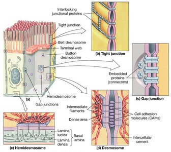
Cell junctions are specialized structures that connect adjacent epithelial cells, providing mechanical strength and communication.
Tight junctions: Seal adjacent cells, preventing passage of substances between them.
Gap junctions: Allow communication via channels formed by connexons, permitting ions and small molecules to pass.
Desmosomes: Anchor cells together, providing resistance to mechanical stress.
Hemidesmosomes: Attach cells to the basement membrane.
Classification of Epithelia
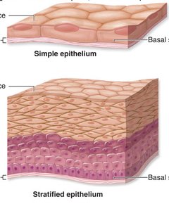
Epithelia are classified based on the number of cell layers and the shape of the cells at the apical surface.
Simple epithelium: One cell layer thick; specialized for absorption, secretion, and filtration.
Stratified epithelium: Multiple layers; provides protection against abrasion.
Pseudostratified epithelium: Appears layered but all cells contact the basement membrane.
Squamous cells: Flat and thin.
Cuboidal cells: Cube-shaped.
Columnar cells: Taller than they are wide.
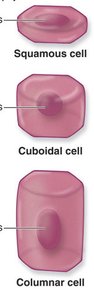
Types of Epithelial Tissue
Simple squamous epithelium: Single layer of flat cells; found in alveoli, lining of blood vessels (endothelium), and serous membranes (mesothelium). Function: rapid diffusion and filtration.

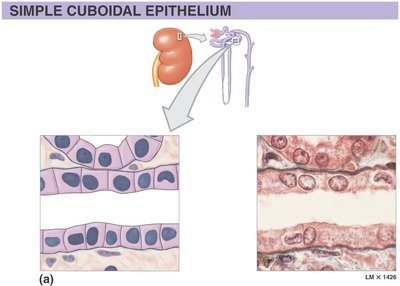
Simple cuboidal epithelium: Single layer of cube-shaped cells; found in kidney tubules, glands, and ducts. Function: secretion and absorption.
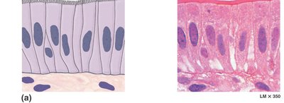
Simple columnar epithelium: Single layer of tall, column-shaped cells; found in the digestive tract. Function: absorption (often with microvilli), secretion, and protection.
Stratified squamous epithelium: Multiple layers of flat cells; found in the epidermis, mouth, esophagus, and vagina. Function: protection against abrasion and pathogens.

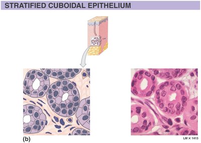
Stratified cuboidal epithelium: Two or more layers of cube-shaped cells; found in sweat gland ducts. Function: protection and secretion.
Pseudostratified ciliated columnar epithelium: Appears layered, but all cells touch the basement membrane; found in the respiratory tract. Function: secretion and movement of mucus by cilia.

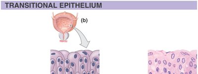
Transitional epithelium: Multiple layers with cells that change shape; found in the urinary bladder. Function: permits stretching and recoil.
Glandular Epithelium
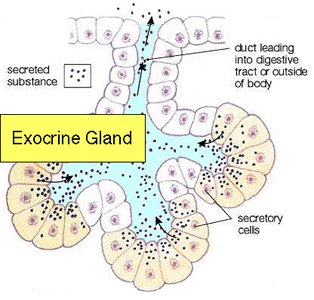
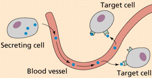
Glands are composed predominantly of epithelial tissue and are specialized for secretion. They are classified as endocrine (secreting into the bloodstream) or exocrine (secreting onto surfaces or into ducts).
Endocrine glands: Release hormones directly into the blood (e.g., thyroid gland).
Exocrine glands: Release secretions through ducts to an epithelial surface (e.g., sweat glands, salivary glands).
Methods of Secretion
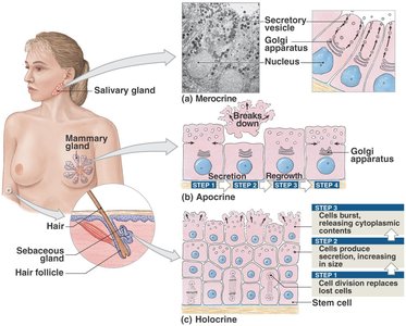
Merocrine: Secretion by exocytosis (e.g., salivary glands, sweat glands).
Apocrine: Apical portion of cell pinches off with secretion (e.g., mammary glands).
Holocrine: Entire cell ruptures to release product (e.g., sebaceous glands).
Connective Tissue
General Characteristics and Functions
Connective tissue is the most abundant and widely distributed tissue type. It supports, binds, and protects other tissues and organs. It consists of cells, protein fibers, and ground substance (matrix).
Cells: Fibroblasts, adipocytes, macrophages, chondrocytes, osteocytes, etc.
Protein fibers: Collagen (strength), elastic (stretch), reticular (supportive networks).
Ground substance: Amorphous material composed of water, proteins, and carbohydrates.
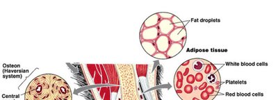
Types of Connective Tissue Proper
Areolar connective tissue: Loose arrangement of fibers and cells; found under epithelia, around blood vessels and nerves. Function: cushions organs, provides support, allows movement.

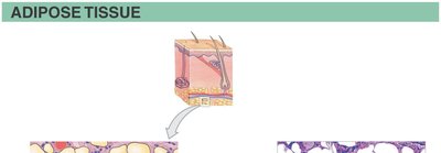
Adipose tissue: Large, closely packed adipocytes; found in hypodermis, around organs. Function: stores energy, insulates, cushions.
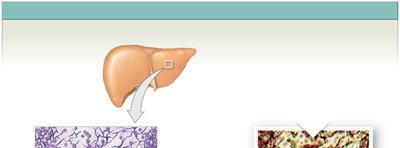
Reticular connective tissue: Network of reticular fibers; found in lymph nodes, spleen, liver. Function: supports cells in soft organs.
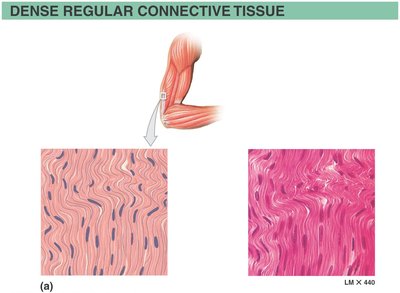
Dense regular connective tissue: Parallel collagen fibers; found in tendons and ligaments. Function: strong attachment, resists pulling forces.
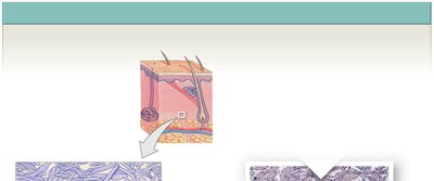
Dense irregular connective tissue: Interwoven collagen fibers; found in dermis, organ capsules. Function: resists tension from multiple directions.
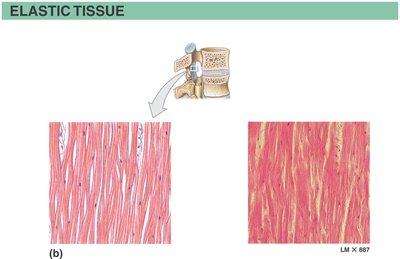
Elastic connective tissue: Abundant elastic fibers; found in walls of arteries, vocal cords. Function: allows stretching and recoil.
Supporting Connective Tissues
Cartilage: Firm, flexible matrix with chondrocytes in lacunae. Types include:
Hyaline cartilage: Most common; found in trachea, nose, ends of long bones.
Elastic cartilage: Flexible; found in ear, epiglottis.
Fibrocartilage: Strongest; found in intervertebral discs, pubic symphysis.
Bone (osseous tissue): Rigid matrix with osteocytes in lacunae; supports, protects, stores minerals, and produces blood cells.
Fluid Connective Tissue
Blood: Composed of red blood cells (erythrocytes), white blood cells (leukocytes), platelets, and plasma. Functions: transport gases, nutrients, wastes, immune response, clotting.
Body Membranes
Types of Body Membranes
Body membranes are composed of epithelial and connective tissues, serving as protective barriers and facilitating various physiological functions.
Mucous membranes: Line passageways open to the exterior (digestive, respiratory tracts); secrete mucus for protection and lubrication.
Serous membranes: Line body cavities not open to the exterior (pleura, pericardium, peritoneum); secrete serous fluid to reduce friction.
Cutaneous membrane: The skin; protects underlying tissues.
Synovial membranes: Line joint cavities; secrete synovial fluid for lubrication.
Muscle Tissue
Types and Functions
Muscle tissue is specialized for contraction, enabling movement of the body and its parts. It is classified into three types:
Skeletal muscle: Voluntary, striated, multinucleated; attached to bones for movement and heat generation.
Cardiac muscle: Involuntary, striated, branched cells with intercalated discs; found in the heart, responsible for pumping blood.
Smooth muscle: Involuntary, non-striated, single nucleus; found in walls of hollow organs and blood vessels, moves substances through tracts.
Nervous Tissue
Structure and Function
Nervous tissue is specialized for the conduction of electrical impulses, allowing rapid communication between body regions. It consists of two main cell types:
Neurons: Generate and transmit electrical signals.
Neuroglia: Support, protect, and nourish neurons.
Locations: Central and peripheral nervous systems.
Summary Table: Characteristics of Major Tissue Types
Tissue Type | Main Function | Key Features | Examples |
|---|---|---|---|
Epithelial | Protection, absorption, secretion | Cellularity, polarity, avascular, regenerative | Skin, lining of GI tract, glands |
Connective | Support, binding, transport | Cells, fibers, ground substance | Tendons, blood, bone, fat |
Muscle | Movement | Contractile cells | Skeletal muscle, heart, GI tract |
Nervous | Communication | Neurons, neuroglia | Brain, spinal cord, nerves |
