 Back
BackChapter 4: Histology – Study Guide for Human Anatomy & Physiology
Study Guide - Smart Notes
 Tailored notes based on your materials, expanded with key definitions, examples, and context.
Histology: The Study of Tissues
Module 4.1 Introduction to Tissues
Histology is the study of tissues, which are groups of structurally and functionally related cells and their external environment, working together to perform common functions. All tissues share two basic components: a discrete population of cells and an extracellular matrix (ECM).
Definition: Histology is the study of normal structures of tissues.
Levels of Organization: Tissues are one level above cells and below organs in the hierarchy of biological organization.
Four Major Tissue Types:
Epithelial tissue: Tightly packed sheets of cells, no visible ECM, covers and lines surfaces, forms glands.
Connective tissue: Connects other tissues, prominent ECM, scattered cells, binds, supports, protects, transports.
Muscle tissue: Generates force by contracting, little ECM.
Nervous tissue: Generates, sends, receives messages, unique ECM.
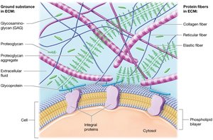
Extracellular Matrix (ECM): Surrounds cells, consists of ground substance and protein fibers, provides strength, directs cell positioning, regulates cell activity.
Cell Junctions: Tight junctions, desmosomes, and gap junctions unite cells in a tissue.
The Extracellular Matrix (ECM)
Ground Substance and Protein Fibers
The ECM is composed of ground substance and protein fibers, which provide structural and functional support to tissues.
Ground Substance: Extracellular fluid with water, nutrients, ions, and macromolecules (GAGs, proteoglycans, glycoproteins).
Glycosaminoglycans (GAGs): Polysaccharide chains (e.g., chondroitin sulfate, hyaluronic acid) attract ions and water, resist compression.
Proteoglycans: GAGs bound to protein core, form aggregates, make ECM firm, resist compression, barrier to diffusion.
Cell-adhesion molecules (CAMs): Glycoproteins that adhere cells to each other and to ECM, maintain tissue architecture.
Protein Fibers:
Collagen fibers: Resist tension and pressure, most abundant protein.
Elastic fibers: Allow extensibility and elasticity.
Reticular fibers: Form meshwork, support cells and ground substance.
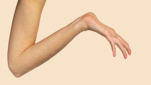
Marfan Syndrome
Marfan syndrome is a genetic disorder affecting the distribution and anchoring of elastic fibers in the ECM, leading to skeletal, cardiovascular, and ocular abnormalities.
Symptoms: Tall stature, long limbs, joint dislocations, heart valve and lens abnormalities, aortic dilation.
Aortic Dissection: Most lethal complication; rupture of aorta can be fatal.

Cell Junctions
Cell junctions are specialized structures that connect cells within tissues, providing structural integrity and communication.
Tight Junctions: Impermeable, lock plasma membranes together, prevent movement of macromolecules.
Desmosomes: Linker proteins attached to intermediate filaments, reinforce structure in tissues under stress.
Gap Junctions: Protein channels allow small substances to flow between cells, important for electrical communication.
Module 4.2 Epithelial Tissues
Functions and Locations
Epithelial tissues cover internal and external surfaces, form barriers, and are involved in protection, secretion, transport, and sensation.
Immune Defense: Physical barriers, immune cells scattered throughout.
Secretion: Form glands producing hormones, oils, mucus.
Transport: Selectively permeable membranes, allow substances to cross by passive or active transport.
Protection: Shield underlying tissues from injury.
Sensation: Rich nerve supply, detect environmental changes.
Components and Classification of Epithelia
Epithelial tissues are tightly packed, avascular, and anchored to underlying connective tissue by a basement membrane (basal lamina and reticular lamina).
Classification: Based on number of cell layers and cell shape.
Simple epithelia: Single cell layer.
Stratified epithelia: Multiple cell layers.
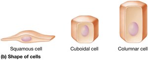
Cell shapes: Squamous (flattened), cuboidal (short), columnar (tall).
Types of Simple Epithelia
Simple squamous epithelium: Thin, rapid diffusion, found in lungs, kidneys, blood vessels.
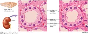
Simple cuboidal epithelium: Cube-shaped, rapid diffusion, found in renal tubules, glands.
Simple columnar epithelium: Rectangular, often with microvilli or cilia, found in intestines, uterine tubes.
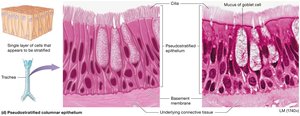
Pseudostratified columnar epithelium: Appears layered, ciliated, found in respiratory tract.
Transport Across Epithelia
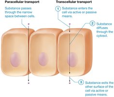
Substances cross simple epithelia via paracellular (between cells) or transcellular (through cells) routes.
Paracellular transport: Limited by tight junctions.
Transcellular transport: Substance enters, diffuses, and exits through cell.
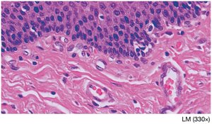
Types of Stratified Epithelia
Keratinized stratified squamous epithelium: Dead, keratin-filled cells, tough, found in skin.
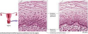
Nonkeratinized stratified squamous epithelium: Alive, moist, found in mouth, esophagus, vagina.
Stratified cuboidal epithelium: Rare, lines sweat gland ducts.
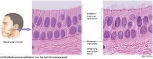
Stratified columnar epithelium: Rare, found in male urethra, cornea, salivary glands.
Transitional epithelium: Only in urinary system, stretches as bladder fills.
Glandular Epithelia
Glands are structures of epithelial origin that synthesize and secrete products. They are classified by shape and method of secretion.
Endocrine glands: Secrete hormones directly into bloodstream.
Exocrine glands: Release products onto surfaces or into ducts.
Goblet cells: Unicellular exocrine glands, secrete mucus.
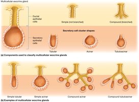
Multicellular glands: Classified by duct structure (simple or compound) and secretory cell shape (tubular, acinar, tubuloacinar).
Methods of Exocrine Secretion
Merocrine: Products released by exocytosis (salivary, sweat glands).
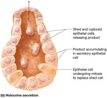
Holocrine: Products released when cell ruptures and dies (sebaceous glands).
Apocrine: Portions of cytoplasm pinched off with product (mammary glands).
Carcinogens and Epithelial Tissues
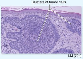
Epithelia are prone to injury and cancer (carcinomas). Basement membrane acts as a barrier to cancer spread.
Examples: Lung adenocarcinoma, breast carcinoma, basal cell carcinoma.
Premalignant: Cancers not yet invaded other tissues.
Module 4.3 Connective Tissues
Types and Functions
Connective tissues are divided into connective tissue proper and specialized connective tissue. They connect, support, protect, and transport substances.
Connective tissue proper: Widely distributed, connects tissues and organs.
Specialized connective tissue: Includes cartilage, bone, and blood.
Cells of Connective Tissue Proper
Adipocytes: Fat cells, store lipids.
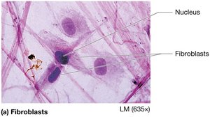
Fibroblasts: Produce protein fibers and ground substance.
Mast cells: Immune cells, release inflammatory mediators.
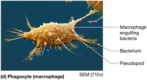
Phagocytes: Macrophages and neutrophils, ingest foreign substances.
Types of Connective Tissue Proper
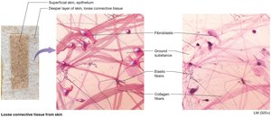
Loose connective tissue: Mostly ground substance, supports blood vessels, houses immune cells.
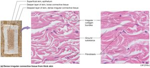
Dense irregular connective tissue: Disorganized collagen bundles, resists tension in all directions.
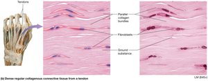
Dense regular connective tissue: Parallel collagen bundles, found in tendons and ligaments.
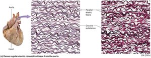
Dense regular elastic connective tissue: Parallel elastic fibers, found in large blood vessels.
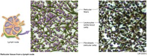
Reticular tissue: Reticular fibers, supports small structures, found in lymph nodes and spleen.
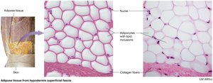
Adipose tissue: Fat-storing cells, insulation, energy reserve, protection.

White and Brown Adipose Tissue
White adipose tissue: One large lipid inclusion, subcutaneous fat, surrounds organs.
Brown adipose tissue: Multiple lipid inclusions, numerous mitochondria, produces heat.
Specialized Connective Tissues
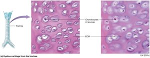
Cartilage: Tough, flexible, absorbs shock, avascular, chondroblasts and chondrocytes.
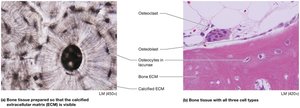
Bone: Hard, supports body, stores calcium, houses marrow, osteoblasts, osteocytes, osteoclasts.
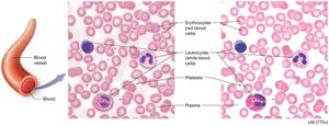
Blood: Fluid ECM (plasma), transports substances, immunity, clotting.
Types of Cartilage
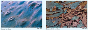
Hyaline cartilage: Most abundant, glossy appearance, found in joints, nose, respiratory tract.
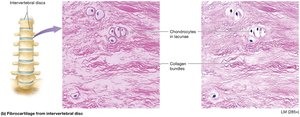
Fibrocartilage: Bundles of collagen, tensile strength, found in intervertebral discs.
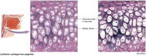
Elastic cartilage: Elastic fibers, vibrates, found in ear and larynx.
Bone Tissue
ECM: 35% organic (collagen, osteoid), 65% inorganic (calcium phosphate).
Cells: Osteoblasts (build), osteocytes (maintain), osteoclasts (destroy).
Osteoarthritis
Osteoarthritis is the degeneration of hyaline cartilage in joints, leading to painful bone-on-bone contact.
Blood
Plasma: Fluid ECM, transports substances.
Erythrocytes: Red blood cells, transport oxygen.
Leukocytes: White blood cells, immunity.
Platelets: Cell fragments, clotting.
Module 4.4 Muscle Tissues
Types and Functions
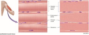
Muscle tissue is specialized for contraction and movement. It consists of excitable muscle cells (myocytes) and a small amount of ECM (endomysium).
Skeletal muscle: Striated, voluntary, multinucleated, attached to skeleton.
Cardiac muscle: Striated, involuntary, uninucleate, found in heart, intercalated discs.
Smooth muscle: Non-striated, involuntary, found in walls of hollow organs.
Module 4.5 Nervous Tissues
Structure and Function
Nervous tissue makes up the brain, spinal cord, and nerves. It consists of neurons and neuroglial cells, with a unique ECM.
Neurons: Excitable, do not divide, consist of cell body (soma), axon (transmits impulses), dendrites (receive impulses).
Neuroglial cells: Support neurons, anchor, monitor fluid, speed transmission, divide by mitosis.
Module 4.7 Membranes
Types and Functions
Membranes are thin sheets of tissue that line body surfaces or cavities. They anchor organs, serve as barriers, function in immunity, and secrete substances.
True membranes: Serous and synovial membranes.
Membrane-like structures: Mucous and cutaneous membranes.
Serous Membranes
Structure: Mesothelium (simple squamous epithelium), basement membrane, connective tissue.
Function: Produce serous fluid, reduce friction, line body cavities.
Synovial Membranes
Structure: Two connective tissue layers, synoviocytes secrete synovial fluid.
Function: Lubricate joints.
Mucous and Cutaneous Membranes
Mucous membranes: Line passages open to outside, produce mucus, protect.
Cutaneous membrane: Skin, keratinized stratified squamous epithelium, protective.
Module 4.8 Tissue Repair
Regeneration and Fibrosis
Tissue repair involves regeneration (replacement with new cells) or fibrosis (replacement with scar tissue).
Regeneration: Epithelial tissues, connective tissue proper, bone, blood.
Fibrosis: Cartilage, cardiac and skeletal muscle, nervous tissue.
Factors affecting repair: Protein supply, vitamin C, blood supply.
Additional info: These notes provide a comprehensive overview of histology, including tissue types, structure, function, and repair, suitable for exam preparation in an anatomy and physiology college course.
