 Back
BackChapter 4: Histology – The Study of Tissues
Study Guide - Smart Notes
 Tailored notes based on your materials, expanded with key definitions, examples, and context.
Histology: The Study of Tissues
Introduction to Tissues
Histology is the study of tissues, which are groups of structurally and functionally related cells and their external environment working together to perform common functions.
All tissues share two basic components: a discrete population of cells related in structure and function, and the surrounding material known as the extracellular matrix (ECM) (ECM differs in composition in each tissue type)
Tissue: Group of related cells and their ECM performing a common function.
Histology: Study of normal tissue structure.
Extracellular Matrix (ECM): Provides strength, directs cell placement, regulates cell activity, and holds cells in position.
Types of Tissues
Epithelial Tissues: Sheets of tightly packed cells with little ECM; cover and line surfaces and form glands.
ex: skin is epithelial tissue
Connective Tissues: Bind, support, protect, and allow transport; cells are scattered in the ECM.
largest group of tissues
ex: your blood is connective tissue because it consists of specialized cells suspended in the ECM, bones are connective tissue because it provides structure, support, & protection for the body
Muscular Tissues: Cells contract to generate force; little ECM.
Nervous Tissues: Neurons generate, send, and receive messages; supported by neuroglial cells with a unique ECM.
nervous tissue forms the brain, spinal cord, & nerves (body's control & communication center). It contains two main cell types: neurons (the electronically active cells that process & transmit information) & neuroglia (the supportive cells that maintain the environment & protect neurons)
Extracellular Matrix (ECM)
Components and Functions
The ECM is composed of substances surrounding the cells in a tissue, providing strength, directing cell placement within a tissue, regulating development, and holding cells in position. It consists of ground substance and protein fibers.
It aids in the survival of cells and helps with mitotic activity
Ground Substance: Gel-like, contains extracellular fluid (ECF), water, ions, nutrients, and macromolecules.
Protein Fibers: Provide tensile strength; include collagen, elastic, and reticular fibers.
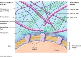
Ground Substance
Glycosaminoglycans (GAGs): Negatively charged polysaccharides (e.g., chondroitin sulfate, hyaluronic acid, these are sugar molecules) that attract positively charged ions in the ECF, drawing water into the ECM.
Proteoglycans: GAGs bonded to protein core; bind to other GAGs & form aggregates, making ECM firmer and resisting compression. Acts as a barrier to diffusion of substances through the ECM
Glycoproteins (Cell-Adhesion Molecules, CAMs): Bind cell surface proteins and fibers, maintaining tissue architecture.
Protein Fibers
Collagen Fibers: Most abundant protein in the body (20 different types of collagen fibers minimum are made in the body); strong and resistant to tension and pressure.
resemble entwined pieces of a steel cable
Elastic Fibers: Made of elastin protein surrounded by glycoproteins; they allow tissues to stretch and return to their original shape.
may stretch to 1/2 time their resting
Distensibility: the length it stretches to without breaking
Elasticity: their original length
Reticular Fibers: Collagen fiber that is thinner than average; interweaves to form a scaffold that supports the cells & ground substance of tissues
forms "webs" in some organs to trap foreign cells
Marfan Syndrome
Results from defects in the gene that codes for glycoprotein fibrillin-1
W/ defective fibrillin-1, elastic fibers cannot function due to improper anchorage in the ECM
Signs & symptoms: tall, long fingers & limbs, skeletal abnormalities, joint dislocations, heart valve abnormalities, & lenses of eyes
Most lethal sign: dilation of the aorta - leads to aortic rupture & fatal blood loss
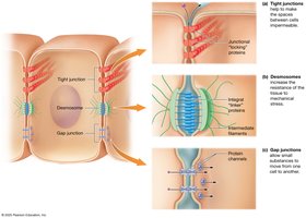
Cell Junctions
Types of Cell Junctions
Cell junctions are connections between neighboring cells, formed by integral proteins.
Tight (Occluding) Junctions: composed of integral "locking" proteins adjacent to plasma membranes; prevent the passage of macromolecules - think of a zipper
They seal cells, control what passes through cells, and protect the body
Ex: Intestines - your intestinal lining absorbs nutrients. W/o tight junctions, stomach acid & bacteria could leak into the surrounding tissues & blood stream
They're made up of proteins like claudins & occludins
Desmosomes: composed of integral "linker" proteins in adjacent plasma membranes; distribute mechanical stress & act like a button
Their main job is to prevent cells from pulling apart when tissue is under stress/stressing
How they work: each cell has proteins that stick out & connect to proteins from the neighboring cell. Inside the cell, these proteins attach to strong protein fibers called intermediate filaments (ex. keratin). The pulling force is distributed across many cells instead of ripping one apart
Ex: skin and heart muscle
Gap Junctions: Protein channels allowing small substances to pass between cells in adjacent plasma membranes
Gap junctions allow the cells to communicate directly by allowing ions/electrical signals to pass quickly between cells
Found in cardiac muscle, smooth muscle
Think "tiny tunnels"
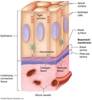
Epithelial Tissues
Epithelial Tissue: found on every external & internal body surface, it acts as a barrier between the body & the environment.
Epithelial tissue is avascular, meaning it has no blood vessels.
Functions of Epithelial Tissue
Protection: Shields underlying tissue from mechanical & thermal injury; produces hard protein keratin; undergoes mitosis rapidly and frequently
Immune Defense: Immune system cells are scattered throughout the tissue
Secretion: Forms glands producing sweat, oil, and hormones.
Transport: Selectively permeable barriers allowing certain substances to pass by passive or active transport from tissue to tissue
Sensation: Supplied with nerves for detecting changes in the internal & external environment; specialized epithelial cells are responsible for some sensations
When Viewing Tissues...
Tissues are thin, stained slices of organs mounted on microscope slides
Due to chemicals, dots & squiggly lines on histological sections "look pink"
When identifying sections, look for visible cells & the chemicals of the ECM that are present
Ex: cells can be identified bc their DNA in the nuclei stains dark purple
Ex: ECM has a ground substance that looks clear or lightly colored, and the protein fibers appear wavy or as straight lines
Refer to Chapter 4 ppt, slides 14-16 & watch the video for further explanation)
Components and Classification of Epithelia
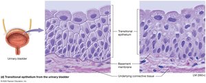
Basement Membrane: Anchors epithelium to connective tissue; consists of basal lamina (epithelial ECM) and reticular lamina (connective tissue ECM).
The basement membrane is a thin layer that sits underneath epithelial tissue to support and anchor it
It acts as a filter (ex, the kidney basement membrane filters blood during urine formation)
Basal Lamina: the ECM of the epithelial tissue consists of collagen fibers & ground substance
Sits directly under the epithelium, "base" of the epithelium
Contains proteins like laminin & collagen
Reticular Lamina: manufactured by the connective tissue deep to the epithelial tissues; consists of reticular fibers & ground substance
Helps attach the basement membrane to connective tissue while adding extra structural support
Contains reticular fibers (primarily collagen fibers)
Cell Surfaces
Apical (free): top/free surface of the epithelial cell; faces the body exterior or the inside of a cavity
Ex: inside of intestines, surface of skin
Lateral (side): the sides of the cell touching neighboring cells; communication, support, cell-to-cell connections
Ex: found in tight junctions, desmosomes, gap junctions
Basal (attached): bottom of the cell, attached to the basement membrane; anchors the cell, allowing nutrients to diffuse from the connective tissue
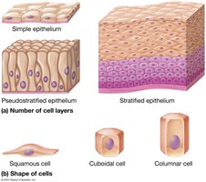
Classification by Layers and Shape
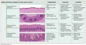
Simple Epithelia: Single cell layer.
Stratified Epithelia: Multiple cell layers.
Pseudostratified Epithelia: Single layer appearing multilayered.
Shape of the Cells
Squamous: Flattened cells.
Cuboidal: Cube-shaped cells.
Columnar: Tall, rectangular cells.
Covering and Lining Epithelia
found on inner & outer body surfaces; form broad, flat sheets of varying thickness (membranes)
Simple Epithelia - one cell layer (thick), does not aid in protection; lines hollow organs & surfaces where diffusion/transport occurs...
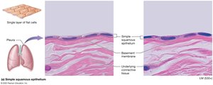
Simple Squamous: Single flat layer (resembles fried eggs fitting together)
functions for rapid diffusion across cells
found in lungs, serous membranes, and blood vessels.
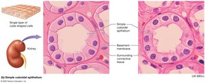
Simple Cuboidal: Single cube-shaped layers
Functions for absorption and secretion
Found in kidney tubules, glands
Appears square w/ a large, central nucleus
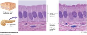
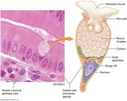
Simple Columnar: Single tall layer of cells & appears rectangular in a section
functions for absorption, secretion, & protection
may have microvilli (tiny finger-like folds on the apical surface) or cilia (longer, hair-like projections
Found in the intestines and uterine tubes.
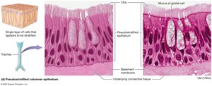
Pseudostratified Columnar: Appears stratified, but is a single layer (nuclei are at different heights & some cells are shorter
often ciliated with goblet cells (cilia are the tiny moving hair-like projections, and they move mucus, which is secreted from goblet cells)
found in the respiratory tract
Step by step respiratory defense system: goblet cells make mucus, dust/bacteria get trapped, cilia move mucus upward, you cough or swallow it - this protects the lungs
Transport Across Simple Epithelia
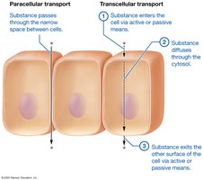
Paracellular Transport: Substances leak between cells.
Transcellular Transport: Substances move through cells
Substance enters through the phospholipid bilayer
Substance diffuses through the cytosol
Substance exits through the other surface of the cell
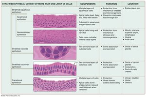
Stratified Epithelia: thicker than simple epithelia; effective protective barriers, common in areas of high stress.
Cell shape changes throughout the thickness of the tissue.
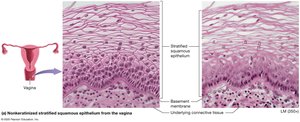
Stratified Squamous (Nonkeratinized): contains multiple layers, has distinct nucleated cells on the apical surface (top/free surface)
found in epithelium of mouth, pharynx, esophagus, anus, and vagina
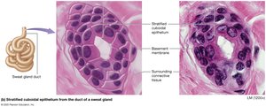
Stratified Cuboidal: two layers of cuboidal cells
lines the ducts of sweaty glands
rare in the body because most body surfaces do not need multiple layers of cube-shaped cells
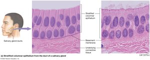
Stratified Columnar: few layers of cells that are columnar in apical layers & cuboidal in basal layers
found in ducts of salivary glands, parts of the male urethra, and the conjunctiva (membrane lining the surface of the eye)
Transitional: cells in the basal layer are cuboidal, and apical cells are dome-shaped when the tissue is relaxed
When stretched, apical cells appear squamous
found in kidney lining, ureters, urinary bladder, and urethra
Summary Tables of Epithelial Tissues
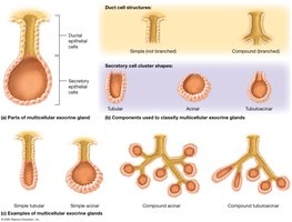
Glandular Epithelia
Gland: Structure that makes and secretes a product; arises from epithelial tissue that grow inward into underlying connective tissue
Glands Release Their Products by 2 Mechanisms
Exocrine Glands: Secrete via ducts lined with epithelial cells to apical surfaces; local action (e.g., sweat, saliva).
Endocrine Glands: Secrete hormones directly into blood; lack ducts; distant action (covered in Ch. 16).
Unicellular Exocrine Glands: Goblet cells secrete mucus in digestive and respiratory tracts.
Multicellular Exocrine Glands: Classified by duct structure (simple/compound) and secretory shape (tubular/acinar/tubuloacinar).
Duct Structure
Simple Glands: ducts do not branch
Compound Glands: ducts do have branches
Shape of Secretory Cell Clusters
Tubular: long and straight or coiled
Acinar: spherical
Tubuloacinar: both tubular & acinar portions
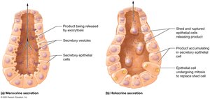
Modes of Secretion
Merocrine: glands package products into secretory vesicles for release by exocytosis (cells stay intact while it secretes product) (e.g., sweat, salivary glands).
Holocrine: Product accumulates in cytosol, which is released when the cell ruptures and dies (e.g., sebaceous glands).
Apocrine: Portion of cytoplasm pinched off with product (e.g., mammary glands and milk droplets).
Connective Tissues
Functions and Classification
Connecting and Binding: Bind tissue layers and anchor organs.
Support: Bone and cartilage support body weight.
Protection: Bone, cartilage, and fat protect organs; immune cells defend against pathogens.
Transport: Blood transports substances throughout the body.
Connective Tissue Proper: Loose, dense, reticular, and adipose tissues.
Specialized Connective Tissues: Cartilage, bone, and blood.
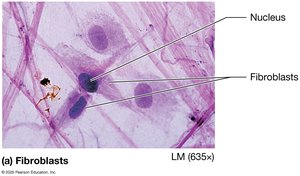
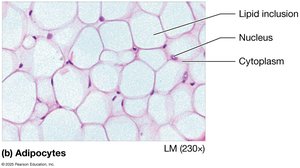
Cells of Connective Tissue Proper
Fibroblasts: Produce fibers and ground substance.
Adipocytes: Fat cells storing lipids.
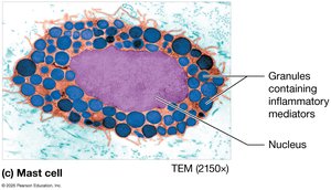
Mast Cells: Release inflammatory mediators (e.g., histamine).
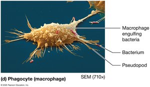
Phagocytes: Engulf foreign substances (e.g., macrophages, neutrophils).
Types of Connective Tissue Proper
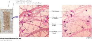
Loose (Areolar) Connective Tissue: All three fiber types, supports epithelium, houses blood vessels.
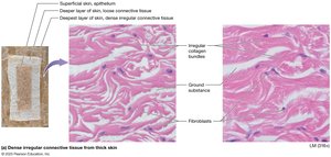
Dense Irregular Connective Tissue: Collagen fibers in random directions; resists tension in all planes; found in dermis.
Dense Regular Collagenous Connective Tissue: Parallel collagen bundles; found in tendons and ligaments.

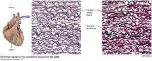
Dense Regular Elastic Connective Tissue: Parallel elastic fibers; found in large blood vessels and some ligaments.
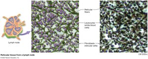
Reticular Tissue: Reticular fibers form supportive networks; found in lymph nodes, spleen.
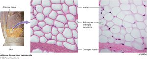
Adipose Tissue: Fat storage, insulation, protection; white and brown types.
Specialized Connective Tissues
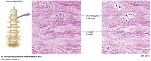
Cartilage
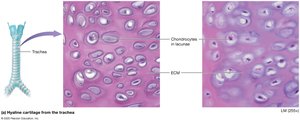
Hyaline Cartilage: Fine collagen; found in joints, nose, respiratory tract, fetal skeleton.
Fibrocartilage: Bundles of collagen; found in intervertebral discs, articular discs.
Additional info:
Elastic cartilage and bone are also discussed in the full chapter, but images were not provided in the selection above. Blood is described as a connective tissue with a fluid ECM (plasma) and cellular components (erythrocytes, leukocytes, platelets).
