 Back
BackChapter 4: Histology – The Study of Tissues
Study Guide - Smart Notes
 Tailored notes based on your materials, expanded with key definitions, examples, and context.
Histology: The Study of Tissues
Introduction to Tissues
Histology is the branch of anatomy that studies tissues, which are groups of structurally and functionally related cells and their surrounding extracellular matrix (ECM). Tissues work together to perform specific functions in the body.
Tissue: A group of related cells and their external environment performing common functions.
Histology: The study of the normal structure of tissues.
All tissues share two basic components: a population of cells and the ECM.
Primary Tissue Types
Overview of the Four Primary Tissue Types
Epithelial Tissues: Sheets of tightly packed cells with little ECM; cover and line body surfaces and cavities; form parts of glands.
Connective Tissues: Connect all other tissues together; cells are scattered through the ECM; bind, support, protect, and allow transport of substances.
Muscular Tissues: Cells contract and generate force; little ECM.
Nervous Tissues: Neurons generate, send, and receive messages; includes supporting cells.
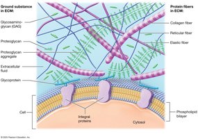
Extracellular Matrix (ECM)
Structure and Function of the ECM
The ECM is composed of substances surrounding the cells in a tissue. It provides strength, directs cell placement, regulates cell development and survival, and holds cells in position. The ECM consists of ground substance and protein fibers.
Ground Substance: Gel-like material containing extracellular fluid, water, ions, nutrients, and solutes.
Protein Fibers: Provide tensile strength. Types include:
Collagen fibers: Resist tension and pressure.
Elastic fibers: Made of elastin; stretch and return to original length.
Reticular fibers: Thinner, shorter collagen fibers.
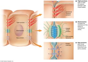
Cell Junctions
Types of Cell Junctions
Cell junctions are connections between neighboring cells, formed by integral proteins. They maintain tissue integrity and regulate movement of substances.
Tight (Occluding) Junctions: Locking proteins prevent passage of macromolecules.
Desmosomes: Linker proteins distribute mechanical stress.
Gap Junctions: Protein channels allow small substances to pass freely between cells.
Epithelial Tissues
Functions of Epithelial Tissue
Epithelial tissue covers all body surfaces, acting as a barrier and performing several key functions:
Protection: Shields underlying tissues from injury; produces keratin; rapid mitosis.
Immune Defense: Contains immune cells.
Secretion: Forms glands that produce substances like sweat and hormones.
Transport: Selectively permeable barriers for substance movement.
Sensation: Innervated to detect environmental changes.
Components and Classification of Epithelia
Cellularity: Densely packed cells, little ECM.
Polarity: Apical (top) and basal (bottom) surfaces.
Basement Membrane: Supports epithelium, connects to underlying connective tissue.
Avascular but Innervated: No direct blood supply, but contains nerves.

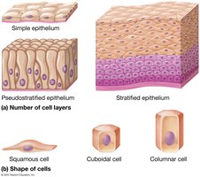
Classification of Epithelia
By Layers:
Simple: One cell layer.
Stratified: Multiple layers.
Pseudostratified: Appears multilayered but is a single layer.
By Cell Shape:
Squamous: Flattened cells.
Cuboidal: Cube-shaped cells.
Columnar: Tall, elongated cells.
Covering and Lining Epithelia
These epithelia are found on inner and outer body surfaces and are often called membranes. Simple epithelia are specialized for absorption, secretion, and filtration, while stratified epithelia provide protection.
Simple Epithelia
Simple Squamous: Single layer of flat cells; found in air sacs of lungs, serous membranes, and blood vessels; allows diffusion and filtration.
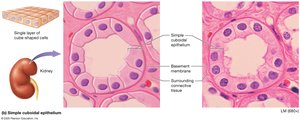
Simple Cuboidal: Single layer of cube-shaped cells; found in kidney tubules and glands; functions in secretion and absorption.
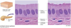
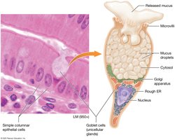
Simple Columnar: Single layer of tall cells; lines digestive tract and uterine tubes; functions in absorption and secretion.
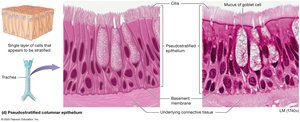
Pseudostratified Columnar: Appears multilayered; found in respiratory tract; secretes and propels mucus.
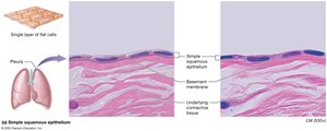
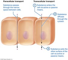
Transport Across Simple Epithelia
Paracellular Transport: Substances leak between cells.
Transcellular Transport: Substances move through cells, entering via the phospholipid bilayer, diffusing through the cytosol, and exiting the other side.
Stratified Epithelia
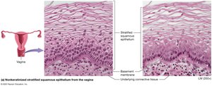
Stratified Squamous: Multiple layers; keratinized (skin) or nonkeratinized (oral mucosa, esophagus, vagina); protects against abrasion.
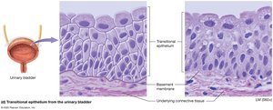
Transitional: Multiple layers; apical cells rounded; found in urinary tract; stretches to allow filling of bladder.
Summary Tables of Epithelial Tissues
Type | Components | Function | Location |
|---|---|---|---|
Simple Squamous | Single layer, flat cells | Diffusion, filtration | Air sacs, blood vessels |
Simple Cuboidal | Single layer, cube cells | Secretion, absorption | Kidney tubules, glands |
Simple Columnar | Single layer, tall cells | Absorption, secretion | Digestive tract, uterus |
Pseudostratified Columnar | Single layer, appears stratified | Secretes, propels mucus | Respiratory tract |
Stratified Squamous | Multiple layers, flat apical cells | Protection | Skin, mouth, esophagus |
Transitional | Multiple layers, rounded apical cells | Stretching | Urinary bladder |
Glandular Epithelia
Glands are structures that make and secrete products. They arise from epithelial tissue and are classified as exocrine or endocrine based on their mode of secretion.
Exocrine Glands: Release products onto surfaces via ducts; local action.
Endocrine Glands: Secrete hormones directly into the blood; lack ducts; act on distant targets.
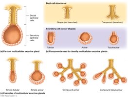
Types of Exocrine Glands
Unicellular: Goblet cells secrete mucus.
Multicellular: Classified by duct structure (simple or compound) and shape (tubular, acinar, tubuloacinar).
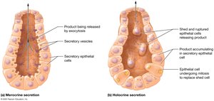
Modes of Secretion
Merocrine: Products released by exocytosis (e.g., salivary, sweat glands).
Holocrine: Cells rupture to release product (e.g., sebaceous glands).
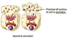
Apocrine: Portion of cytoplasm pinched off with product (e.g., mammary glands).
Connective Tissues
Functions of Connective Tissue
Connecting and Binding: Bind tissue layers and anchor organs.
Support: Bone and cartilage support body weight.
Protection: Bone protects organs; cartilage and fat absorb shock.
Transport: Blood transports substances throughout the body.
Classification of Connective Tissue
Connective Tissue Proper: Loose, dense, reticular, and adipose tissues; widely distributed.
Specialized Connective Tissues: Cartilage, bone, and blood.
Cells of Connective Tissue Proper
Fibroblasts: Produce fibers and ground substance.
Adipocytes: Store fat.
Mast Cells: Immune cells with granules.
Phagocytes: Engulf foreign substances (e.g., macrophages, neutrophils).
Types of Connective Tissue Proper
Loose (Areolar) Connective Tissue: Loosely arranged fibers; binds epithelia to other tissues; allows passage of nerves and vessels.
Dense Irregular Connective Tissue: Densely packed collagen in random directions; resists tension in multiple directions.
Dense Regular Collagenous Connective Tissue: Parallel collagen fibers; found in tendons and ligaments; resists tension in one direction.
Dense Regular Elastic Connective Tissue: Parallel elastic fibers; allows organs to stretch.
Reticular Tissue: Reticular fibers; forms framework for lymphatic organs.
Adipose Tissue: Fat tissue; insulates, protects, stores energy.
Specialized Connective Tissues
Cartilage: Tough, flexible; chondrocytes in lacunae; types include hyaline, fibrocartilage, and elastic cartilage.
Bone: Supports, protects, stores calcium; contains osteoblasts, osteocytes, osteoclasts; compact and spongy types.
Blood: Fluid ECM (plasma); contains erythrocytes, leukocytes, and platelets; transports substances and provides immunity.
Muscle Tissues
Types of Muscle Tissue
Skeletal Muscle: Long, cylindrical, multinucleated, striated; voluntary movement.
Cardiac Muscle: Branching, uninucleated, striated, intercalated discs; involuntary contractions in the heart.
Smooth Muscle: Spindle-shaped, single nucleus, no striations; involuntary movement in hollow organs and blood vessels.
Nervous Tissue
Structure and Function
Neurons: Generate, conduct, and receive nerve impulses; consist of cell body, axon, and dendrites; amitotic.
Neuroglial Cells: Support neurons, anchor blood vessels, monitor ECF, speed impulse transmission, circulate CSF; can divide by mitosis.
Tissues in Organs
Integration of Tissues
Organs are composed of two or more tissue types working together. For example, skeletal muscle contains muscle and connective tissue, while the trachea contains epithelial, connective, cartilage, and muscle tissues.
Membranes
Types of Membranes
Serous Membranes: Line body cavities; produce serous fluid for lubrication.
Synovial Membranes: Line joint cavities; secrete synovial fluid for lubrication.
Mucous Membranes: Line passages opening to the exterior; secrete mucus.
Cutaneous Membrane: The skin; covers the body surface.
Tissue Repair
Regeneration and Fibrosis
Regeneration: Damaged cells replaced by the same type; common in epithelial and most connective tissues.
Fibrosis: Fibroblasts produce collagen to fill defects, resulting in scar tissue; occurs in cartilage, cardiac, and skeletal muscle, and nervous tissue.
Capacity for Repair
Epithelial Tissues: High regenerative capacity due to stem cells.
Connective Tissues: Most regenerate well; cartilage heals by fibrosis.
Smooth Muscle: Usually regenerates.
Cardiac and Skeletal Muscle: Heal by fibrosis due to limited mitosis.
Nervous Tissue: Neurons generally do not regenerate.
