 Back
BackChapter 4: Histology – The Study of Tissues
Study Guide - Smart Notes
 Tailored notes based on your materials, expanded with key definitions, examples, and context.
Histology: The Study of Tissues
Introduction to Tissues
Histology is the study of tissues, which are groups of structurally and functionally related cells and their surrounding extracellular matrix (ECM). Tissues perform common functions and are a key level of organization in the human body, situated between cells and organs.
Tissue: A group of similar cells and their ECM performing a specific function.
All tissues share two basic components: a population of cells and the ECM.
Types of Tissues
There are four primary tissue types, each defined by their cellular composition, ECM, and function:
Epithelial tissue: Tightly packed sheets of cells with little to no visible ECM. They cover and line body surfaces and cavities, and form glands.
Connective tissue: Connects other tissues, with abundant ECM and scattered cells. Functions include binding, support, protection, and transport.
Muscle tissue: Specialized for contraction and force generation, with little ECM.
Nervous tissue: Specialized for generating, sending, and receiving electrical messages, with unique ECM.
The Extracellular Matrix (ECM)
Components and Functions
The ECM is a complex network of substances surrounding cells, providing structural and biochemical support. It consists of ground substance and protein fibers.
Ground substance: Extracellular fluid with water, nutrients, ions, and macromolecules (glycosaminoglycans, proteoglycans, glycoproteins).
Protein fibers: Collagen (strength), elastic (stretch and recoil), and reticular (supportive meshwork).
Functions: Provides strength, directs cell positioning, regulates development and survival.
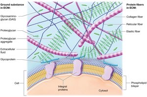
Ground Substance Details
Glycosaminoglycans (GAGs): Polysaccharide chains that attract ions and water, helping ECM resist compression.
Proteoglycans: GAGs bound to protein cores, forming aggregates that make ECM firm and act as diffusion barriers.
Cell-adhesion molecules (CAMs): Glycoproteins that anchor cells to each other and the ECM, maintaining tissue structure.
Protein Fibers in ECM
Collagen fibers: Provide tensile strength and resistance to stretching.
Elastic fibers: Allow tissues to stretch and return to original shape.
Reticular fibers: Form supportive networks in organs like the spleen.
Clinical Connection: Marfan Syndrome
Genetic disorder affecting elastic fiber distribution in ECM.
Symptoms: Tall stature, long limbs, joint dislocations, heart and eye abnormalities.
Most serious complication: Aortic dissection (rupture).

Cell Junctions
Tight junctions: Seal cells together, preventing passage of substances between them (e.g., in blood vessels).
Desmosomes: Anchor cells together via intermediate filaments, providing mechanical strength (e.g., skin).
Gap junctions: Protein channels allowing communication and passage of small molecules between cells (e.g., cardiac muscle).
Epithelial Tissues
Functions and Characteristics
Epithelial tissues cover body surfaces, line cavities, and form glands. They are tightly packed, avascular, and rest on a basement membrane.
Functions: Protection, immune defense, secretion, selective transport, sensation.
Basement membrane: Composed of basal lamina (from epithelial cells) and reticular lamina (from connective tissue).
Classification of Epithelia
By layers: Simple (one layer), Stratified (multiple layers).
By shape: Squamous (flat), Cuboidal (cube-shaped), Columnar (tall).
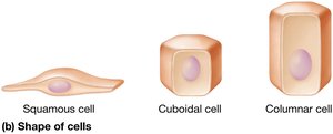
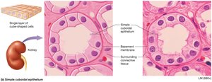
Types of Simple Epithelia
Simple squamous: Thin, rapid diffusion (lungs, blood vessels).
Simple cuboidal: Cube-shaped, secretion/absorption (kidney tubules, glands).
Simple columnar: Tall, absorption/secretion (digestive tract, uterine tubes).
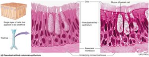
Pseudostratified columnar: Appears layered, but all cells touch basement membrane (respiratory tract).
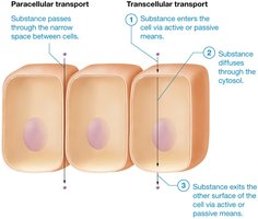
Transport Across Epithelia
Paracellular transport: Substances pass between cells (limited by tight junctions).
Transcellular transport: Substances pass through the cell (via membrane transport).
Types of Stratified Epithelia
Keratinized stratified squamous: Dead, keratin-filled cells at surface (skin).
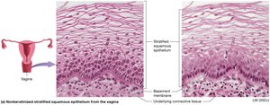
Nonkeratinized stratified squamous: Living cells at surface, moist (mouth, esophagus, vagina).
Stratified cuboidal: Rare, lines sweat gland ducts.
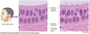
Stratified columnar: Rare, found in male urethra, eye, and some glands.
Transitional epithelium: Urinary system, stretches as bladder fills.
Glandular Epithelia
Glands: Epithelial structures that secrete substances.
Endocrine glands: Secrete hormones into blood (no ducts).
Exocrine glands: Secrete products onto surfaces or into ducts (e.g., sweat, saliva).
Goblet cells: Unicellular exocrine glands that secrete mucus.
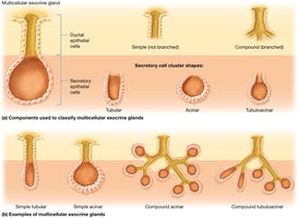
Multicellular exocrine glands classified by duct structure (simple/compound) and secretory shape (tubular, acinar, tubuloacinar).
Methods of Exocrine Secretion
Merocrine: Exocytosis (most glands).
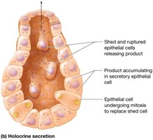
Holocrine: Cell ruptures to release product (sebaceous glands).
Apocrine: Portion of cytoplasm pinched off (rare, mammary glands).
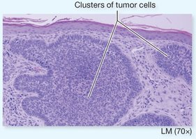
Carcinomas
Carcinoma: Cancer of epithelial tissue (e.g., lung, breast, skin).
Basement membrane acts as a barrier to cancer spread; invasion occurs when cancer cells degrade this barrier.
Connective Tissues
Overview and Functions
Connective tissues support, connect, protect, and transport substances throughout the body. They are characterized by abundant ECM and various cell types.
Types: Connective tissue proper and specialized connective tissue (cartilage, bone, blood).
Functions: Binding, support, protection, transport.
Cells of Connective Tissue Proper
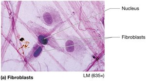
Fibroblasts: Produce fibers and ground substance.
Adipocytes: Store fat.
Mast cells: Release inflammatory mediators.
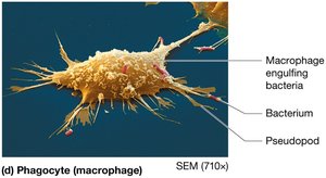
Phagocytes: Engulf foreign material (macrophages, neutrophils).
Types of Connective Tissue Proper
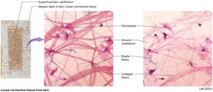
Loose connective tissue (areolar): Supports epithelia, contains all fiber types, houses immune cells.
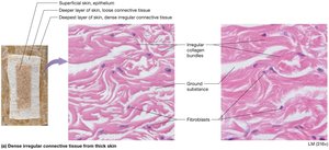
Dense irregular connective tissue: Disorganized collagen bundles, resists tension in multiple directions (dermis).
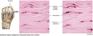
Dense regular connective tissue: Parallel collagen bundles, resists tension in one direction (tendons, ligaments).
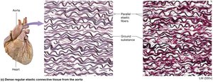
Dense regular elastic connective tissue: Parallel elastic fibers, allows stretch (large blood vessels).
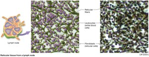
Reticular tissue: Network of reticular fibers, supports lymphatic organs.
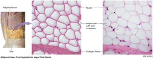
Adipose tissue: Fat storage, insulation, protection.

White adipose: Energy storage, insulation.
Brown adipose: Heat production, more mitochondria.
Specialized Connective Tissues
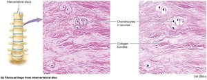
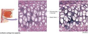
Cartilage: Flexible, shock-absorbing, avascular. Types: hyaline, fibrocartilage, elastic.
Bone: Rigid, supports body, stores calcium, houses marrow.
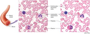
Blood: Fluid ECM (plasma), transports cells and substances.
Types of Cartilage
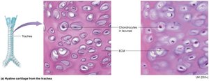
Hyaline cartilage: Most common, glassy ECM, found in joints, nose, respiratory tract.
Fibrocartilage: Dense collagen bundles, strong, found in intervertebral discs.
Elastic cartilage: Abundant elastic fibers, flexible, found in ear and epiglottis.
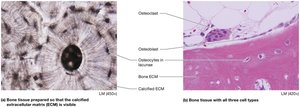
Bone Tissue
ECM: 35% organic (collagen, osteoid), 65% inorganic (calcium phosphate).
Cells: Osteoblasts (build), osteocytes (maintain), osteoclasts (resorb).
Blood
ECM: Plasma (fluid, proteins).
Cells: Erythrocytes (O2 transport), leukocytes (immunity), platelets (clotting).
Muscle Tissues
Types and Functions
Muscle tissue is specialized for contraction and movement. It contains excitable cells called myocytes and a small amount of ECM (endomysium).
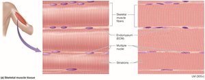
Skeletal muscle: Striated, voluntary, multinucleated, attached to bones.
Cardiac muscle: Striated, involuntary, branched, one nucleus, found in heart, connected by intercalated discs.
Smooth muscle: Non-striated, involuntary, spindle-shaped, found in walls of hollow organs.
Nervous Tissues
Structure and Function
Nervous tissue is found in the brain, spinal cord, and nerves. It consists of neurons and neuroglial cells, with a unique ECM.
Neurons: Excitable cells with a cell body (soma), axon (sends impulses), and dendrites (receive impulses).
Neuroglial cells: Support, protect, and assist neurons; capable of mitosis.
Membranes
Types and Functions
Membranes are thin sheets of tissue that line body surfaces or cavities. They anchor organs, serve as barriers, and secrete substances.
Serous membranes: Line body cavities, produce serous fluid to reduce friction.
Synovial membranes: Line joint cavities, secrete synovial fluid for lubrication.
Mucous membranes: Line passages open to the outside, secrete mucus for protection.
Cutaneous membrane: The skin, protective barrier.
Tissue Repair
Regeneration and Fibrosis
Regeneration: Replacement of dead/damaged cells with the same cell type, restoring function (common in epithelial and most connective tissues).
Fibrosis: Replacement with dense irregular connective tissue (scar tissue), resulting in loss of function (common in cardiac, skeletal muscle, and nervous tissue).
Repair depends on cell mitotic ability, protein and vitamin C availability, and blood supply.
