 Back
BackChapter 4: Histology – The Study of Tissues
Study Guide - Smart Notes
 Tailored notes based on your materials, expanded with key definitions, examples, and context.
Introduction to Tissues
Overview of Tissues
Tissues are groups of similar cells that perform a common function. The human body contains approximately 50 trillion cells, organized into four primary tissue types: epithelial, connective, nervous, and muscular tissue. The study of tissues and their organization into organs is called histology (microscopic anatomy).
Epithelial tissue: Covers surfaces and lines cavities.
Connective tissue: Supports, binds, and protects organs.
Nervous tissue: Specialized for communication.
Muscular tissue: Specialized for contraction and movement.
Epithelial Tissue
General Characteristics
Epithelial tissue forms the external and internal linings of many organs and body surfaces. It consists of closely packed cells with minimal extracellular material, is avascular (lacks blood vessels), but is innervated (supplied by nerves), and is capable of rapid regeneration. Epithelial cells rest on a basement membrane that anchors them to underlying connective tissue.
Basal surface: Faces the basement membrane.
Apical surface: Faces away from the basement membrane, toward the body surface or cavity.
Classification of Epithelial Tissue
Epithelial tissues are classified by the number of cell layers and the shape of the cells at the apical surface.
Simple epithelium: One cell layer thick.
Stratified epithelium: Multiple layers of cells.
Cell shapes: Squamous (flat), cuboidal (cube-shaped), columnar (tall and column-like).
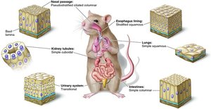
Types of Epithelial Tissue
Simple Squamous Epithelium
Function: Rapid diffusion and filtration; secretion of serous fluid.
Location: Lung alveoli, kidney glomeruli, inner lining of blood vessels (endothelium), serous membranes.
Simple Cuboidal Epithelium
Function: Secretion.
Location: Most glands (liver, thyroid, mammary, salivary), kidney tubules.
Simple Columnar Epithelium
Function: Absorption; secretion of mucus (by goblet cells); may have cilia or microvilli.
Location: Lining of stomach and intestines.
Pseudostratified Columnar Epithelium
Function: Secretes and propels mucus; appears multilayered but all cells touch the basement membrane.
Location: Respiratory tract.
Stratified Squamous Epithelium
Function: Protection against abrasion; deepest layers undergo continuous mitosis.
Types: Keratinized (skin surface); Nonkeratinized (tongue, oral mucosa, esophagus, vagina, anus).
Transitional Epithelium
Function: Allows for stretching; surface cells change from round to flat when stretched.
Location: Ureter and bladder.
Connective Tissue
General Characteristics
Connective tissue is the most abundant and widely distributed tissue type. It is highly vascular (except cartilage), with cells separated by abundant extracellular matrix. The matrix consists of fibers and ground substance.
Cells: Fibroblasts, adipocytes, osteocytes, chondrocytes, blood cells, mast cells, macrophages.
Matrix: Extracellular material composed of fibers and ground substance.
Connective Tissue Fibers
Collagenous fibers: Tough, flexible, resist stretching; most abundant protein in the body.
Reticular fibers: Thin collagen fibers coated with glycoprotein; form supportive networks.
Elastic fibers: Made of elastin; allow tissues to stretch and recoil.
Ground Substance
The ground substance is a clear, colorless, viscous material that fills the spaces between cells and fibers, slowing the movement of pathogens and facilitating nutrient and waste exchange.
Functions of Connective Tissue
Binding of organs (tendons, ligaments)
Support (bones, cartilage)
Physical protection (cranium, ribs, sternum)
Immune protection (white blood cells)
Movement (bones as levers)
Storage (fat, calcium, phosphorus)
Heat production (brown fat in infants)
Transport (blood)
Types of Connective Tissue
Loose Connective Tissue
Areolar tissue: Loosely organized fibers, abundant ground substance; underlies epithelia, in serous membranes, between muscles.
Reticular tissue: Mesh of reticular fibers and fibroblasts; forms supportive stroma for lymphatic organs (lymph nodes, spleen, thymus, bone marrow).
Dense Connective Tissue
Dense regular: Densely packed, parallel collagen fibers; found in tendons and ligaments.
Dense irregular: Densely packed, randomly arranged collagen fibers; found in deeper skin layers and organ capsules.
Adipose Tissue
Composed of adipocytes; stores energy, insulates, cushions organs.
Cartilage
Supportive, flexible matrix; avascular; types include hyaline, elastic, and fibrocartilage.
Hyaline cartilage: Articular surfaces, costal cartilage, trachea, fetal skeleton.
Elastic cartilage: External ear, epiglottis.
Fibrocartilage: Pubic symphysis, intervertebral discs, menisci.
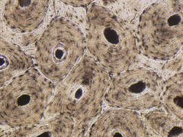
Bone
Rigid matrix with osteocytes in lacunae; structural unit is the osteon.
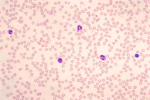
Blood
Fluid connective tissue; transports cells and dissolved substances.
Plasma: Liquid ground substance.
Formed elements: Erythrocytes (red blood cells), leukocytes (white blood cells), platelets (cell fragments).
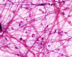
Areolar Tissue
Areolar tissue is a type of loose connective tissue with loosely organized fibers and abundant ground substance. It underlies all epithelia, is found in serous membranes, and between muscles.
Nervous Tissue
Structure and Function
Nervous tissue is specialized for communication by electrical and chemical signals. It consists of neurons, which detect stimuli, respond quickly, and transmit information, and neuroglia (glial cells), which support and protect neurons.
Muscular Tissue
Types of Muscle Tissue
Skeletal muscle: Voluntary, striated, multinucleated, attached to bones.
Cardiac muscle: Involuntary, striated, intercalated discs, highly fatigue resistant, found in the heart.
Smooth muscle: Involuntary, non-striated, tapered cells, forms sheets in organs.
Cell Junctions
Types of Cell Junctions
Tight junctions: Seal adjacent cells together, preventing passage of substances between them.
Desmosomes: Patch-like junctions that hold cells together, providing mechanical strength.
Gap junctions: Channels that allow ions and small molecules to pass between cells, enabling communication.
Glands
Types of Glands
Endocrine glands: Ductless; secrete hormones directly into the blood (e.g., thyroid, adrenal, pituitary).
Exocrine glands: Secrete substances onto body surfaces or into cavities via ducts (e.g., sweat, mammary, tear glands).
Types of Secretions
Serous glands: Produce thin, watery secretions (e.g., perspiration, milk, tears, digestive juices).
Mucous glands: Produce mucin, which forms mucus when hydrated; goblet cells are unicellular mucous glands.
Cytogenic glands: Release whole cells (e.g., sperm and egg cells).
Modes of Secretion
Merocrine: Secretion by exocytosis (e.g., tear glands, pancreas, sweat glands).
Apocrine: Secretion by budding off a portion of the cell (e.g., mammary glands for milk fat).
Holocrine: Secretion by cell disintegration, releasing cell fragments and products (e.g., oil glands of scalp and skin).
Summary Table: Epithelial Tissue Types and Locations
Type | Function | Location |
|---|---|---|
Simple Squamous | Diffusion, filtration | Lung alveoli, blood vessels, serous membranes |
Simple Cuboidal | Secretion | Kidney tubules, glands |
Simple Columnar | Absorption, secretion | Intestines, stomach |
Pseudostratified Columnar | Secretion, propulsion of mucus | Respiratory tract |
Stratified Squamous | Protection | Skin, oral cavity, esophagus, vagina, anus |
Transitional | Stretching | Urinary bladder, ureter |
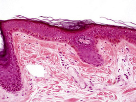
Example: Histological Images
Blood: Shows erythrocytes (red blood cells) and leukocytes (white blood cells).
Stratified Squamous Epithelium: Multiple layers, protection against abrasion.
Bone Tissue: Osteons with central canals and concentric lamellae.
Areolar Connective Tissue: Loose arrangement of fibers and cells.
Additional info: The images provided reinforce the microscopic appearance of key tissue types discussed in this chapter, aiding in visual identification for laboratory and exam settings.
