 Back
BackChapter 4: Skin and Body Membranes – Study Notes
Study Guide - Smart Notes
 Tailored notes based on your materials, expanded with key definitions, examples, and context.
Body Membranes
Classification and Function of Body Membranes
Body membranes are thin layers of tissue that cover surfaces, line body cavities, and form protective sheets around organs. They are classified based on their tissue composition and location in the body.
Epithelial membranes: Include cutaneous, mucous, and serous membranes.
Connective tissue membranes: Include synovial membranes.
Functions:
Cover body surfaces
Line body cavities
Form protective sheets around organs
Epithelial Membranes
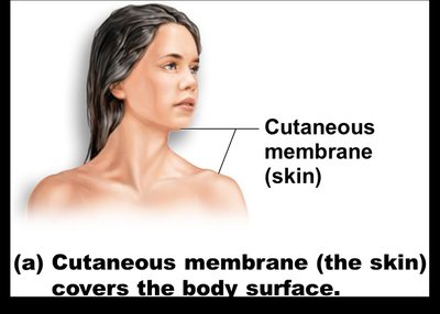
Cutaneous Membrane (Skin)
The cutaneous membrane, commonly known as the skin, is the outermost protective boundary of the body. It is a dry membrane exposed to air and consists of a superficial epidermis (keratinized stratified squamous epithelium) and an underlying dermis (dense connective tissue).
Function: Protection from environmental hazards, dehydration, and pathogens.
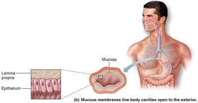
Mucous Membranes
Mucous membranes line all body cavities that open to the exterior, such as the respiratory, digestive, urinary, and reproductive tracts. The type of surface epithelium depends on the site (e.g., stratified squamous in the mouth, simple columnar in the digestive tract). The epithelium rests on a loose connective tissue layer called the lamina propria.
Function: Adapted for absorption or secretion; protects underlying tissues.
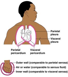
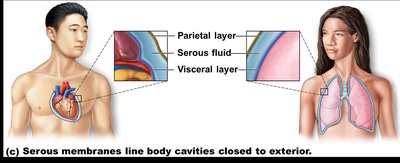
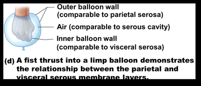
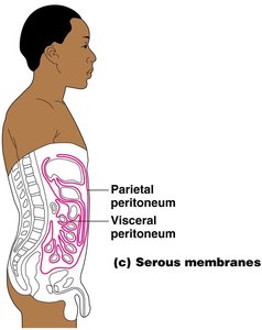
Serous Membranes
Serous membranes line body cavities that are closed to the exterior. They consist of a surface layer of simple squamous epithelium and an underlying thin layer of areolar connective tissue. Serous membranes occur in pairs separated by serous fluid: the visceral layer covers organs, and the parietal layer lines the cavity wall.
Function: Reduce friction between organs and cavity walls by secreting serous fluid.
Examples: Peritoneum (abdominal cavity), pleura (lungs), pericardium (heart).
Connective Tissue Membranes
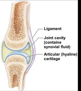
Synovial Membranes
Synovial membranes are composed entirely of connective tissue and contain no epithelial cells. They line the fibrous capsules surrounding joints and tendon sheaths, secreting a lubricating synovial fluid for smooth joint movement.
Function: Reduce friction in joints and nourish articular cartilage.
The Integumentary System (Skin)
Components of the Integumentary System
The integumentary system consists of the skin (cutaneous membrane) and its derivatives, including sweat glands, oil glands, hair, and nails. It serves as the body's first line of defense and plays a vital role in homeostasis.
Functions of the Integumentary System
The skin performs several essential functions for the body:
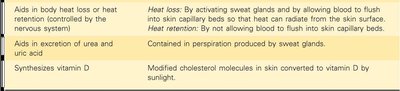
Function | How Accomplished |
|---|---|
Protection | Mechanical, chemical, bacterial, and thermal barrier; prevents desiccation and UV damage. |
Aids in body heat loss or heat retention | Heat loss: Activates sweat glands and increases blood flow to skin capillaries. Heat retention: Reduces blood flow to skin capillaries. |
Aids in excretion of urea and uric acid | Contained in perspiration produced by sweat glands. |
Synthesizes vitamin D | Modified cholesterol molecules in skin are converted to vitamin D by sunlight. |

Structure of the Skin
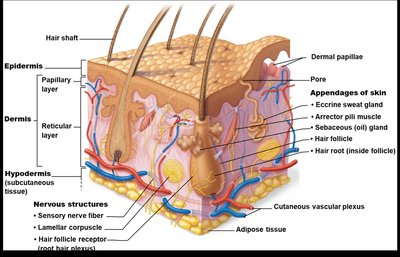
The skin is composed of three main layers: the epidermis, dermis, and subcutaneous tissue (hypodermis).
Epidermis: Outermost layer, made of keratinized stratified squamous epithelium.
Dermis: Dense connective tissue beneath the epidermis.
Subcutaneous tissue (hypodermis): Deep to the dermis, primarily adipose tissue; anchors skin to underlying organs, absorbs shock, and insulates.
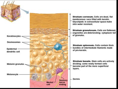
Layers of the Epidermis (Deep to Superficial: BSGLC)
Stratum basale (germinativum): Deepest layer; cells undergo mitosis and move upward.
Stratum spinosum: Cells contain thick bundles of intermediate filaments.
Stratum granulosum: Cells flatten, organelles deteriorate, cytoplasm full of granules.
Stratum lucidum: Present only in thick, hairless skin (palms, soles); dead cells.
Stratum corneum: Outermost layer; shingle-like dead cells filled with keratin.
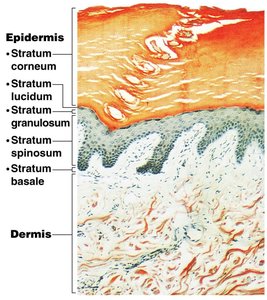
Dermis
The dermis consists of two layers:
Papillary layer: Upper region with dermal papillae, capillary loops, and sensory receptors.
Reticular layer: Deepest skin layer; contains blood vessels, sweat and oil glands, and deep pressure receptors.
Collagen fibers: Provide toughness.
Elastic fibers: Provide elasticity.
Blood vessels: Help regulate body temperature.
Skin Pigment and Colour Determinants
Skin color is determined by three main pigments:
Melanin: Produced by melanocytes in the stratum basale; yellow, brown, or black pigment; amount depends on genetics and sunlight exposure.
Carotene: Orange-yellow pigment from some vegetables.
Hemoglobin: Red coloring from blood cells in dermal capillaries; oxygen content affects redness.
Appendages of the Skin
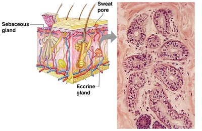
Cutaneous Glands
Sebaceous (oil) glands: Produce sebum, a lubricant that keeps skin soft, prevents brittle hair, and kills bacteria. Most ducts empty into hair follicles; some open directly onto the skin surface. Activated at puberty.
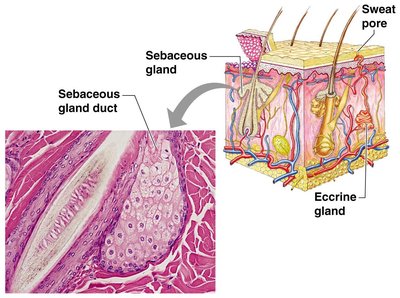
Sweat (sudoriferous) glands: Two types:
Eccrine glands: Open via duct to pore on skin surface; produce sweat (mostly water, salts, vitamin C, metabolic waste).
Apocrine glands: Ducts empty into hair follicles; produce sweat with fatty acids and proteins (odor from bacteria).
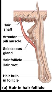
Hair and Hair Follicles
Hair follicle: Dermal and epidermal sheath surrounds hair root.
Arrector pili muscle: Smooth muscle that pulls hairs upright when cold or frightened.
Sebaceous and sweat glands: Associated with hair follicles.
Homeostatic Imbalances of Skin
Burns
Burns are tissue damage and cell death caused by heat, electricity, UV radiation, or chemicals. Major dangers include dehydration, electrolyte imbalance, and circulatory shock.
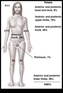
Rule of Nines
The body is divided into 11 areas, each representing about 9% of total body surface area, to quickly estimate the extent of burns.
Severity of Burns
First-degree: Only epidermis damaged; skin is red and swollen.
Second-degree: Epidermis and upper dermis damaged; skin is red with blisters.
Third-degree: Destroys entire skin layer; burn is gray-white or black.
Burns are considered critical if:
Over 25% of body has second-degree burns
Over 10% of body has third-degree burns
There are third-degree burns of the face, hands, or feet
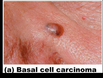
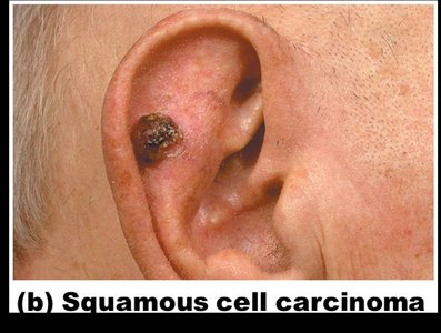
Skin Cancer
Skin cancer is the most common type of cancer and is classified as benign (does not spread) or malignant (metastasizes to other parts of the body).
Basal cell carcinoma: Least malignant, most common; arises from stratum basale.
Squamous cell carcinoma: Can metastasize to lymph nodes; arises from stratum spinosum; believed to be sun-induced.
Malignant melanoma: Most deadly; cancer of melanocytes; metastasizes rapidly to lymph and blood vessels.
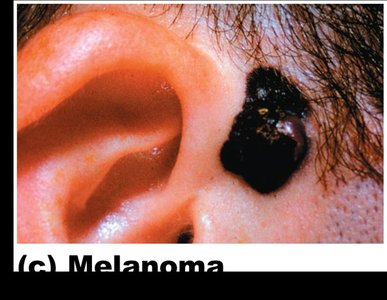
ABCD Rule for Detecting Melanoma
A = Asymmetry: Two sides of pigmented mole do not match.
B = Border irregularity: Borders are not smooth.
C = Color: Different colors in pigmented area.
D = Diameter: Spot is larger than 6 mm.
