 Back
BackChapter 4: The Tissue Level of Organization – Comprehensive Study Notes
Study Guide - Smart Notes
 Tailored notes based on your materials, expanded with key definitions, examples, and context.
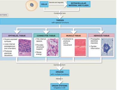
Tissues: An Overview
Definition and Classification
Tissues are groups of closely associated cells that perform related functions and share similar structural characteristics. The human body contains four primary types of tissues, each with distinct roles:
Epithelium – coverings and linings
Connective tissue – support
Nervous tissue – control
Muscle tissue – movement
Tissues are composed of cells suspended in an extracellular matrix, which consists of proteins and interstitial fluid. The composition and distribution of cells and matrix vary by tissue type.
Epithelial Tissue
Functions and Characteristics
Epithelial tissues cover body surfaces and line body cavities. All substances entering or leaving the body must pass through an epithelium. Key functions include:
Protection of underlying tissues
Absorption, secretion, and ion transport
Filtration of molecules from fluid
Formation of slippery surfaces within the body
Epithelial tissue contains little or no extracellular matrix.
Key Characteristics of Epithelial Tissue
High Cellularity: Cells are separated by minimal extracellular matrix and are connected by specialized junctions.
Polarity: The apical (exposed) surface differs structurally and functionally from the basal (attached) surface.
Regeneration: Lost cells are rapidly replaced by cell division.
Avascular: Epithelia lack blood vessels and receive nutrients via diffusion from underlying connective tissue. However, they are innervated.
Attachment: Epithelial sheets are supported and attached to underlying connective tissue by a basal membrane.
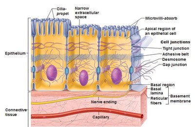
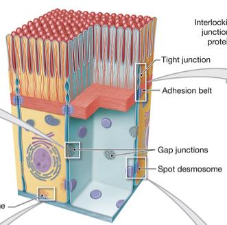
Lateral Cell Junctions
Specialized attachment sites, known as cell junctions, connect epithelial cells to each other or to the extracellular matrix. The three main types are:
Tight junctions: Fusion of plasma membranes prevents diffusion of fluids and solutes between cells.
Desmosomes: Anchoring junctions that reinforce tight junctions and prevent tearing. Spot desmosomes form small discs; hemidesmosomes attach cells to the basement membrane.
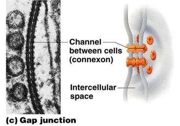
Gap junctions: Connexons form narrow passages for direct movement of small molecules between cells, facilitating intercellular communication.


Naming and Classification of Epithelial Tissues
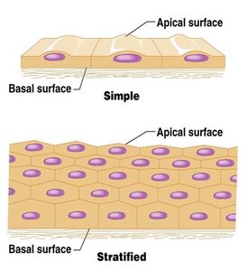
Epithelial tissues are named based on the number of cell layers and the shape of the cells:
Simple: One layer of cells covering the basement membrane.
Stratified: More than one layer of cells covering the basement membrane.
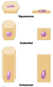
The second word describes cell shape:
Squamous: Cells are wider than tall.
Cuboidal: Cells are as wide as tall, like cubes.
Columnar: Cells are taller than they are wide, like columns.
Types of Epithelial Tissues
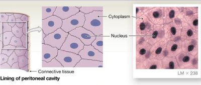
Simple Squamous Epithelium
Single layer of flat cells with disc-shaped nuclei. Functions include reducing friction, controlling permeability, and secretion in serous membranes. Found in renal corpuscles, alveoli of lungs, serous membranes, and blood vessels.
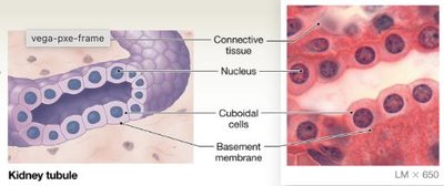
Simple Cuboidal Epithelium
Single layer of cube-like cells with large, spherical central nuclei. Functions include limited protection, secretion, and absorption. Found in kidney tubules, thyroid gland, and secretory portions of some glands.
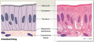
Simple Columnar Epithelium
Single layer of column-shaped cells with oval nuclei. Functions include protection, absorption, and secretion. Found in lining of stomach, intestines, gallbladder, uterine tubes, and small bronchi. May be ciliated or have microvilli.
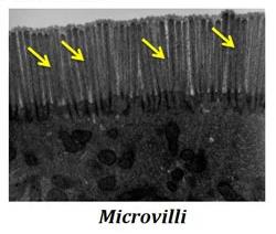
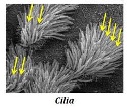
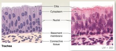
Cilia vs. Microvilli
Cilia: Highly motile extensions that sweep mucus, dust, and pathogens out of airways.
Microvilli: Non-motile extensions that increase surface area for absorption and provide a passive barrier against pathogens.
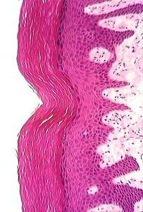
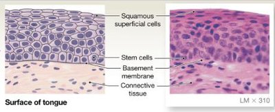
Stratified Squamous Epithelium
Several layers of cells; basal cells are cuboidal or columnar, surface cells are squamous. Types include keratinized (epidermis of skin) and non-keratinized (moist linings of mouth, throat, esophagus, rectum, anus, and vagina). Function: protection.
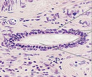
Stratified Cuboidal Epithelium
Generally two layers of cube-shaped cells. Functions: protection. Locations: ducts of mammary glands, salivary glands, and largest sweat glands. Rare tissue.
Stratified Columnar Epithelium
Several layers of cells; basal cells are usually cuboidal, superficial cells are columnar. Functions: protection and secretion. Locations: urethra, portions of pharynx, epiglottis, large ducts of some excretory glands. Rare tissue.
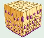
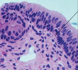
Pseudostratified Columnar Epithelium
Single layer of cells of varying heights, nuclei at different depths. Functions: secretion of mucus, ciliated type propels mucus or reproductive cells. Locations: ducts of male reproductive tubes, lining of trachea and upper respiratory tract, auditory tube, internal ear.
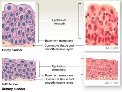
Transitional Epithelium
Cells transition between two shapes; superficial cells appear dome-like when relaxed, flatten out when stretched. Functions: stretches and permits distension, impermeable to urine. Locations: lines ureters, urinary bladder, and urethra.
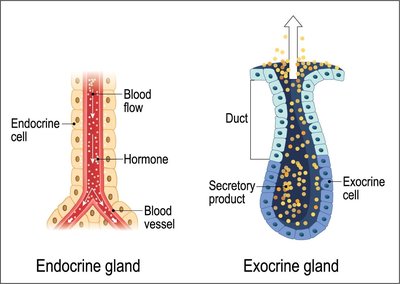
Glandular Epithelium
Many epithelial tissues contain gland cells that produce exocrine or endocrine secretions.
Endocrine glands: Ductless, secrete hormones directly into bloodstream.
Exocrine glands: Ducts carry products to epithelial surface (e.g., mucus, salivary, sweat, oil glands).
Modes of Exocrine Secretion
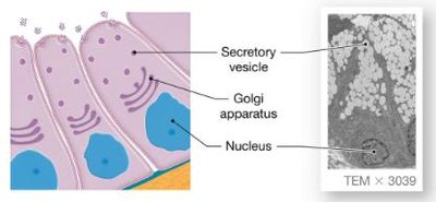
Merocrine: Product released by exocytosis from secretory vesicles at apical surface. Most common method (e.g., salivary glands).
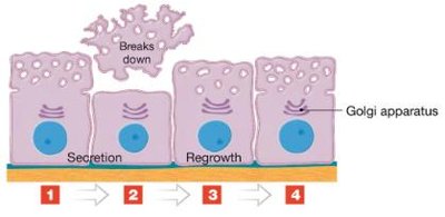
Apocrine: Loss of apical cytoplasm; inclusions and vesicles are shed, cell regrows before releasing more secretions (e.g., mammary glands).
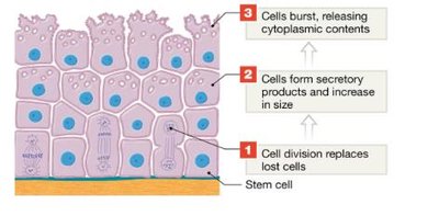
Holocrine: Secretion occurs as superficial gland cell bursts; replacement by division of underlying stem cells (e.g., sebaceous glands).
Gland Morphology
Unicellular glands: Composed of one cell (e.g., goblet cells produce mucus).
Multicellular glands: Composed of multiple cells, with epithelium-walled duct and secretory unit.
Connective Tissue
General Characteristics
Connective tissues are the most diverse and abundant in the human body. All derive from a common embryonic origin called mesenchyme. General features include:
Specialized cells separated by large amounts of extracellular matrix
Extracellular matrix = ground substance + protein fibers
Underlies epithelial tissues
Highly vascularized and innervated
Functions include structural framework, transport, protection, tissue interconnection, fat storage, and defense against microorganisms.
Composition of Connective Tissue
Connective tissue can be described by the following equations:
Specialized Cells in Connective Tissue
Fibroblasts: Always present, produce fibers and ground substance.
Fibrocytes: Maintain connective tissue fibers.
Chondroblasts/Chondrocytes: In cartilage, secrete and maintain matrix.
Osteoblasts/Osteocytes: In bone, secrete and maintain matrix.
Macrophages: Phagocytic cells, mobilize immune system.
Adipocytes: Fat cells, store nutrients.
Mast cells: Promote inflammation, contain histamine and heparin.
Extracellular Matrix
Ground substance: Hydrophilic, varies in consistency (liquid in blood, crystallized in bone), holds tissue fluid.
Protein fibers:
Collagen fibers: Long, straight, unbranched; strongest and most abundant, resist tension.
Reticular fibers: Mesh-like network, provide flexibility and support.
Elastic fibers: Branched, wavy, stretch and recoil.
Types of Connective Tissues
Connective tissues are divided into four broad classes based on ground substance consistency:
Connective tissue proper: Syrupy ground substance
Cartilage: Gelatinous ground substance
Bone: Crystallized ground substance
Blood: Liquid ground substance
Connective Tissue Proper
Characterized by syrupy ground substance, fibroblasts, fibrocytes, defense cells, and adipocytes. Types vary in density and fiber types.
Loose connective tissues: Fibers separated by ground substance
Areolar: Wraps and cushions organs, supports movement, defense against pathogens. Found under epithelia, between muscles, around joints, blood vessels, nerves.
Adipose: Closely packed adipocytes, stores food, insulates, supports organs. Found deep to skin, around kidneys, abdomen, breasts.
Reticular: Network of reticular fibers, forms internal skeleton for lymphoid organs. Found in liver, kidney, lymph nodes, spleen, bone marrow.
Dense connective tissues: Densely packed fibers, little ground substance
Dense regular: Parallel collagen fibers, provides firm attachment, found in tendons, ligaments.
Dense irregular: Irregularly arranged collagen fibers, resists forces from many directions, found in deep dermis, fibrous capsules.
Elastic: Predominantly elastic fibers, allows recoil, found in walls of arteries, bronchial tubes.
Cartilage
Firm, flexible gel matrix with water and protein fibers. Specialized cells: chondroblasts (immature), chondrocytes (mature). Avascular and non-innervated, weakly regenerative. Types:
Hyaline cartilage: Most common, imperceptible collagen fibers, chondrocytes in lacunae. Functions: support, resists stress, reduces friction. Locations: costal cartilage, synovial joints, larynx, trachea, fetal skeleton, ends of long bones.
Elastic cartilage: More elastic fibers, maintains shape, flexibility. Locations: external ear, epiglottis, auditory tube.
Fibrocartilage: Less ground substance, dense collagen fibers, durable and tough. Functions: resists compression, absorbs shock. Locations: intervertebral discs, knee joint, pubic bone.
Bone
Hard, calcified matrix with flexible collagen fibers. Specialized cells: osteoblasts (immature), osteocytes (mature). Well vascularized and innervated, highly regenerative. Types: compact and spongy.
Blood
Atypical connective tissue with cells surrounded by liquid matrix (plasma). Specialized cells: red and white blood cells. Functions: transport of gases, nutrients, wastes. Location: within blood vessels.
Tissue Membranes
Types and Functions
Tissue membranes are physical barriers composed of epithelial tissues supported by connective tissues. They cover or line broad areas and separate spaces within the body. Most are moist and modified for secretion. Four types:
Cutaneous membrane: Skin, covers outer surface
Mucous membrane: Coated with mucus, lines digestive, respiratory, urinary, reproductive tracts
Serous membrane: Lines peritoneal, pleural, pericardial cavities, secretes serous fluid
Synovial membrane: Lines joint cavities, secretes synovial fluid
Serous Membranes
Serous membranes are continuous, with parietal layer lining the cavity wall and visceral layer lying directly on the organ. Examples:
Pleural membranes: Line the lungs
Pericardial membranes: Line the heart
Peritoneal membranes: Line abdominopelvic organs
Muscle Tissue
General Characteristics
Muscle tissue is composed of elongated muscle cells (fibers) specialized to contract. Contraction is due to interaction between myosin and actin filaments. Muscle tissue is highly innervated and vascularized. Three types:
Skeletal muscle tissue: Long, cylindrical, multinucleate, striated. Functions: voluntary movement, stabilizes skeleton, generates heat, protects organs. Location: skeletal muscles.
Cardiac muscle tissue: Branching cells, striated, usually one nucleus, intercalated discs (desmosomes and gap junctions). Functions: involuntary contraction to propel blood, circulates blood, maintains pressure. Location: heart.
Smooth muscle tissue: Short, spindle-shaped, one central nucleus, no striations, forms sheets. Functions: propels substances, involuntary control. Locations: walls of blood vessels, digestive, respiratory, urinary, reproductive organs.
Nervous Tissue
General Characteristics
Nervous tissue contains neurons and neuroglial cells with minimal connective tissue support. Neurons generate and conduct electrical impulses; neuroglia provide support, maintain chemical composition, supply nutrients, and defend against infection.
Neurons: Cell body (nucleus), dendrites (projections), axon (single projection).
Neuroglia: Support cells.
Functions: transmit electrical signals, integrate information, transmit signals to effectors. Locations: brain, spinal cord, nerves.
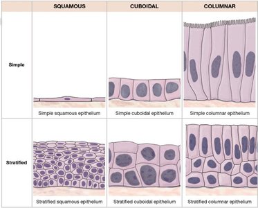
Summary Table: Classification of Epithelial Tissues
Squamous | Cuboidal | Columnar | |
|---|---|---|---|
Simple | Simple squamous epithelium | Simple cuboidal epithelium | Simple columnar epithelium |
Stratified | Stratified squamous epithelium | Stratified cuboidal epithelium | Stratified columnar epithelium |
Additional info: Academic context was added to clarify tissue functions, cell types, and histological features. All images included are directly relevant to the adjacent explanations and reinforce key concepts.
