 Back
BackChapter 4: The Tissue Level of Organization – Study Notes
Study Guide - Smart Notes
 Tailored notes based on your materials, expanded with key definitions, examples, and context.
Chapter 4: The Tissue Level of Organization
Introduction to Tissues
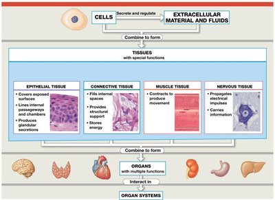
Tissues are groups of specialized cells and cell products that perform specific functions. The study of tissues is called histology. There are four basic types of tissue in the human body: epithelial, connective, muscle, and nervous tissue.
Types of Tissues
Epithelial Tissue: Covers exposed surfaces, lines internal passageways, and forms glands.
Connective Tissue: Fills internal spaces, supports other tissues, transports materials, and stores energy.
Muscle Tissue: Specialized for contraction and movement.
Nervous Tissue: Carries information throughout the body via electrical impulses.
Epithelial Tissue
General Characteristics
Epithelial tissue includes both epithelia (layers of cells covering surfaces) and glands. Epithelia form the surface of the skin and line the digestive, respiratory, reproductive, and urinary tracts—areas exposed to the external environment.
Functions of Epithelial Tissue
Physical Protection: Protects surfaces from abrasion, dehydration, and chemical/biological agents.
Control Permeability: Regulates entry and exit of substances; permeability varies by tissue type.
Sensation: Contains sensory receptors for various senses.
Specialized Secretions: Gland cells discharge secretions for lubrication, protection, temperature regulation, or as chemical messengers.
Structural Characteristics of Epithelia
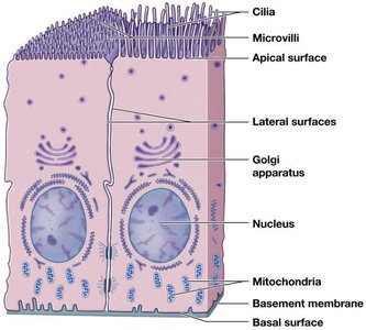
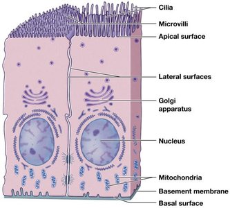
Polarity: Structural and functional differences between apical (exposed) and basal (attached) surfaces.
Cellularity: Cells are tightly bound by cell junctions.
Attachment: Base is bound to a noncellular basement membrane.
Avascularity: No blood vessels; nutrients diffuse or are absorbed across surfaces.
Regeneration: High rate of cell replacement via stem cell division.
Surface Specializations
Microvilli: Small projections that increase surface area for absorption/secretion (e.g., digestive tract).
Cilia: Long extensions that move fluids or secretions over surfaces (e.g., respiratory tract).
Intercellular Connections
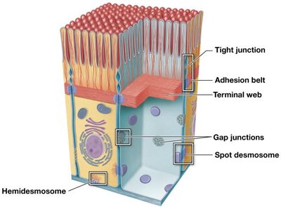
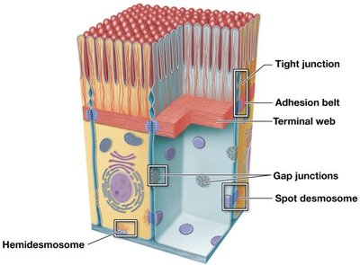
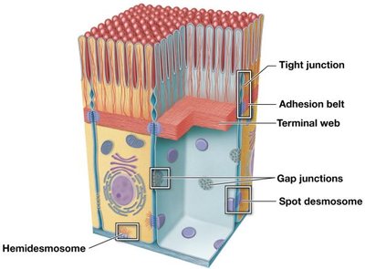
Epithelial cells are firmly attached to each other and to the basement membrane via two main types of connections: cell adhesion molecules (CAMs) and cell junctions.
Cell Junctions
Gap Junctions: Allow communication between cells via connexons (protein channels).
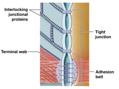
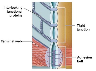
Tight Junctions: Prevent passage of water and solutes between cells; maintain compartmentalization.
Desmosomes: Provide strong attachment between cells; spot desmosomes connect to intermediate filaments, hemidesmosomes attach cells to the basement membrane.
Classification of Epithelia
Epithelia are classified by cell shape and number of layers:
Shapes: Squamous (thin, flat), Cuboidal (box-like), Columnar (tall, slender).
Layers: Simple (one layer), Stratified (multiple layers).
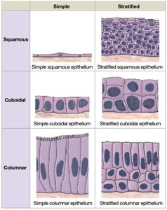
Table: Epithelial Tissue Classification
Simple | Stratified | |
|---|---|---|
Squamous | Simple squamous epithelium | Stratified squamous epithelium |
Cuboidal | Simple cuboidal epithelium | Stratified cuboidal epithelium |
Columnar | Simple columnar epithelium | Stratified columnar epithelium |
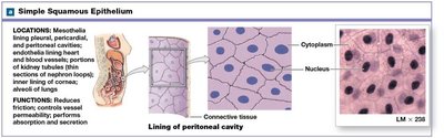
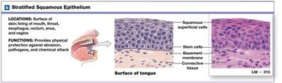
Squamous Epithelium
Simple Squamous Epithelium: Delicate, found in protected regions for absorption/diffusion (e.g., alveoli of lungs, lining of heart and blood vessels—endothelium, lining of body cavities—mesothelium).
Stratified Squamous Epithelium: Located where mechanical stresses are severe (e.g., skin, mouth, esophagus, anus, vagina). Keratinized type is tough and water-resistant (epidermis); nonkeratinized type resists abrasion but must be kept moist.
Cuboidal and Columnar Epithelium
Simple Cuboidal: Limited protection; secretion/absorption (e.g., glands, kidney tubules).
Stratified Cuboidal: Rare; found in ducts of sweat and mammary glands.
Transitional Epithelium: Stratified; tolerates stretching (e.g., urinary bladder).
Simple Columnar: Absorption/secretion (e.g., stomach, intestines).
Pseudostratified Columnar: Appears layered but is single-layered; often ciliated (e.g., respiratory tract).
Stratified Columnar: Rare; found in pharynx, epiglottis, anus, urethra.
Glandular Epithelium
Endocrine Glands: Ductless; secrete hormones into bloodstream (e.g., thyroid, pituitary).
Exocrine Glands: Secrete onto epithelial surfaces or into ducts (e.g., sweat, tears, milk).
Connective Tissue
General Characteristics
Connective tissues consist of specialized cells, extracellular protein fibers, and ground substance. The matrix (fibers + ground substance) makes up most of the tissue volume and surrounds the cells.
Functions of Connective Tissue
Structural framework for the body
Transport of fluids and dissolved materials
Protection of organs
Support, surround, and interconnect other tissues
Energy storage (triglycerides)
Defense against pathogens
Categories of Connective Tissue
Connective Tissue Proper: Loose and dense types; many cell types and fibers.
Fluid Connective Tissues: Blood and lymph; watery matrix with dissolved proteins.
Supporting Connective Tissues: Cartilage and bone; densely packed fibers.
Cells of Connective Tissue Proper
Fibroblasts: Most abundant; secrete hyaluronan and protein subunits for fibers (collagen, reticular, elastic).
Adipocytes: Fat cells; store lipids for energy, insulation, and protection.
Mesenchymal Cells: Stem cells for repair and regeneration.
Melanocytes: Synthesize melanin pigment.
Macrophages: Phagocytic immune cells; fixed or free.
Mast Cells: Release histamine (inflammation) and heparin (anticoagulant).
Lymphocytes: Immune cells; some become plasma cells (antibody production).
Fibers of Connective Tissue Proper
Collagen Fibers: Strong, flexible, unbranched; found in tendons and ligaments.
Reticular Fibers: Thin, branching; form supportive frameworks in organs.
Elastic Fibers: Contain elastin; allow tissues to stretch and return to shape (e.g., elastic ligaments in vertebral column).
Ground Substance
Clear, colorless, viscous substance that fills spaces between cells and fibers; slows pathogen movement.
Types of Connective Tissue Proper
Loose Connective Tissue: "Packing material"; includes areolar, adipose, and reticular tissue.
Dense Connective Tissue: Collagenous; includes dense regular (tendons, ligaments, aponeuroses), dense irregular (dermis, organ capsules), and elastic tissue (walls of large blood vessels).
Fluid Connective Tissue
Blood: Watery matrix (plasma) with formed elements (RBCs, WBCs, platelets).
Lymph: Monitored by lymphocytes; flows through lymphatic vessels.
Supporting Connective Tissue
Cartilage: Firm gel matrix with chondroitin sulfates; cells are chondrocytes in lacunae; avascular; types include hyaline, elastic, and fibrocartilage.
Bone (Osseous Tissue): Calcified matrix (calcium salts + collagen fibers); cells are osteocytes in lacunae; highly vascular; covered by periosteum.
Muscle Tissue
Types of Muscle Tissue
Skeletal Muscle: Long, multinucleate fibers; striated voluntary muscle; limited repair via myosatellite cells.
Cardiac Muscle: Found in heart; connected by intercalated discs; striated involuntary muscle; limited repair.
Smooth Muscle: Found in walls of blood vessels and hollow organs; nonstriated involuntary muscle; capable of regeneration.
Nervous Tissue
General Characteristics
Nervous tissue is specialized for the propagation of electrical impulses. Most nervous tissue is found in the brain and spinal cord. It contains two main cell types: neurons and neuroglia.
Neurons
Cell Body: Contains nucleus and organelles.
Dendrites: Receive information from other neurons.
Axon: Conducts electrical signals to other cells.
Neuroglia
Supporting cells of nervous tissue; several types with various functions.
