 Back
BackChapter 4: The Tissue Level of Organization – Study Notes
Study Guide - Smart Notes
 Tailored notes based on your materials, expanded with key definitions, examples, and context.
The Tissue Level of Organization
Introduction to Tissues
Tissues are collections of specialized cells and cell products that perform specific functions. Organs are composed of two or more types of tissues, and histology is the study of tissue structure. Understanding tissues is fundamental to anatomy and physiology, as they form the basis for organ structure and function.
Tissue: Group of cells with similar structure and function.
Histology: The study of tissues.
Organs: Structures made of multiple tissue types.
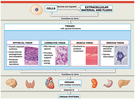
Four Major Types of Tissue
Overview of Tissue Types
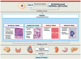
The human body contains four basic types of tissue, each with distinct roles and characteristics:
Epithelial Tissue: Covers exposed surfaces, lines internal passageways, and forms glands.
Connective Tissue: Fills internal spaces, supports other tissues, transports materials, and stores energy.
Muscle Tissue: Specialized for contraction and movement.
Nervous Tissue: Carries electrical signals throughout the body.
Epithelial Tissue
Structure and Functions
Epithelial tissue includes epithelia and glands. Epithelia are layers of cells covering internal or external surfaces, while glands produce secretions. Epithelial tissue serves several essential functions:
Physical Protection: Protects surfaces from abrasion, dehydration, and chemical/biological agents.
Control Permeability: Regulates substances entering or leaving the body.
Provide Sensation: Contains sensory receptors for various senses.
Produce Specialized Secretions: Glands release hormones (endocrine) or other secretions (exocrine).
Characteristics of Epithelia
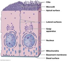
Apical Surface: Exposed surface, may have microvilli (increase absorption/secretion) or cilia (move fluids).
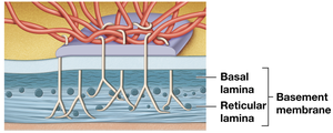
Attachment: Base bound to basement membrane (basal lamina).
Avascularity: No blood vessels; nutrients diffuse from underlying tissues.
Cellularity: Cells tightly bound by cell junctions.
Regeneration: Continuously replaced by stem cell division.
Intercellular Connections
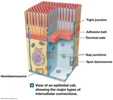
Epithelial cells are connected by specialized junctions that maintain tissue integrity and allow communication:
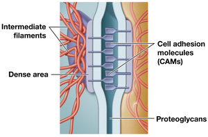
Cell Adhesion Molecules (CAMs): Transmembrane proteins attaching cells to each other and the extracellular matrix.
Proteoglycans: Act as intercellular cement.
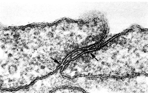
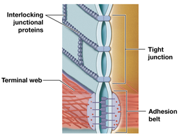
Types of Cell Junctions
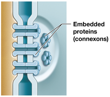
Gap Junctions: Allow passage of ions and small molecules; important for cell communication.
Tight Junctions: Prevent passage of water and solutes between cells.
Desmosomes: Provide strong attachment between cells; connected to cytoskeleton.
Hemidesmosomes: Anchor cells to the basement membrane.
Classification of Epithelia
By Shape and Layers
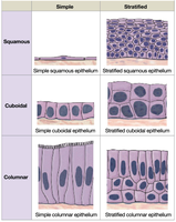
Epithelia are classified based on cell shape and the number of cell layers:
Shapes: Squamous (thin, flat), Cuboidal (boxy), Columnar (tall, slender).
Layers: Simple (single layer), Stratified (multiple layers).
Types of Epithelia
Simple Squamous: Absorption and diffusion; lines lung alveoli, body cavities (mesothelium), heart and blood vessels (endothelium).
Stratified Squamous: Protection against mechanical stress; skin, mouth, anus. Keratinized for strength and water resistance.
Simple Cuboidal: Secretion and absorption; glands, kidney tubules.
Stratified Cuboidal: Rare; ducts of sweat and mammary glands.
Transitional Epithelium: Changes appearance with stretching; lines urinary bladder.
Simple Columnar: Absorption and secretion; stomach, intestines.
Pseudostratified Columnar: Appears layered, but is single; ciliated; nasal cavity, trachea, bronchi.
Stratified Columnar: Rare; protection in pharynx, anus, urethra.
Glandular Epithelia
Endocrine Glands: Release hormones into bloodstream; no ducts.
Exocrine Glands: Discharge secretions onto surfaces through ducts.
Methods of Secretion
Merocrine: Product released by exocytosis (e.g., sweat glands).
Apocrine: Product released by shedding cytoplasm (e.g., mammary glands).
Holocrine: Product released by cell bursting and dying (e.g., sebaceous glands).
Connective Tissue
Structure and Functions
Connective tissues are diverse and provide support, protection, and transport. They consist of specialized cells, extracellular protein fibers, and ground substance (fluid).
Matrix: Extracellular components (protein fibers and ground substance).
Functions: Structural framework, transport, protection, support, energy storage, defense.
Categories of Connective Tissue
Connective Tissue Proper: Viscous matrix, rich in protein fibers; includes loose and dense types.
Fluid Connective Tissues: Watery matrix; blood and lymph.
Supporting Connective Tissues: Densely packed fibers; cartilage and bone.
Connective Tissue Proper
Cells
Fibroblasts: Most abundant; secrete ground substance and fibers.
Adipocytes: Store fat.
Mesenchymal Cells: Stem cells; respond to injury.
Melanocytes: Produce melanin pigment.
Macrophages: Immune cells; engulf pathogens and debris.
Fibers
Collagen: Strong, flexible; found in tendons and ligaments.
Elastic: Stretchy, return to original length.
Reticular: Branching network; supports organs.
Ground Substance
Clear, colorless, viscous; fills spaces and slows pathogen movement.
Loose Connective Tissues
Areolar: Loosely organized; supports epithelia.
Adipose: Contains adipocytes; padding, insulation, energy storage.
Reticular: Rich in reticular fibers; supports organs.
Dense Connective Tissues
Dense Regular: Parallel collagen fibers; tendons, ligaments.
Dense Irregular: Interwoven collagen; dermis, capsules.
Elastic: Mainly elastic fibers; blood vessels, respiratory passages.
Fluid Connective Tissues
Blood and Lymph
Blood: Plasma (matrix), red blood cells, white blood cells, platelets.
Lymph: Forms as interstitial fluid enters lymphatic vessels.
Supporting Connective Tissues
Cartilage and Bone
Cartilage: Firm gel matrix with chondroitin sulfates; chondrocytes in lacunae; avascular.
Types: Hyaline (tough, flexible), Elastic (flexible), Fibrocartilage (durable, tough).
Bone: Calcified matrix with collagen fibers; osteocytes in lacunae; highly vascular; covered by periosteum.
Tissue Membranes
Types and Functions
Mucous Membranes: Line passageways open to exterior; digestive, respiratory, urinary, reproductive tracts.
Serous Membranes: Line internal cavities; reduce friction; parietal and visceral portions.
Cutaneous Membrane: Skin; covers body.
Synovial Membranes: Line joint cavities; produce synovial fluid.
Muscle Tissue
Types and Features
Skeletal Muscle: Long, multinucleate fibers; voluntary movement.
Cardiac Muscle: Short, branched cells; intercalated discs; involuntary, pumps blood.
Smooth Muscle: Spindle-shaped cells; involuntary; walls of hollow organs.
Nervous Tissue
Structure and Function
Neurons: Conduct electrical impulses; cell body, dendrites (input), axon (output).
Neuroglia: Supporting cells.
Tissue Response to Injury
Inflammation and Regeneration
Inflammation: Triggered by trauma or infection; mast cells release chemicals; necrosis and abscess may occur.
Regeneration: Varies by tissue; epithelia, connective, and smooth muscle regenerate well; skeletal, cardiac, and nervous tissue regenerate poorly.
Fibrosis: Replacement by fibrous tissue; does not restore normal function.
Aging and Tissue Structure
Aging Effects
Slows repair and maintenance; tissues become thinner and more fragile.
Increased bruising; decreased regeneration speed and effectiveness.
Cancer Incidence
Cancer rates increase with age; majority caused by environmental factors.
