 Back
BackChapter 4: Tissue – The Living Fabric (Study Notes)
Study Guide - Smart Notes
 Tailored notes based on your materials, expanded with key definitions, examples, and context.
Tissue: The Living Fabric
Introduction to Tissues
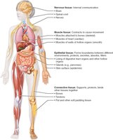
Tissues are groups of cells with similar structure and function, essential for maintaining homeostasis in the human body. The study of tissues is known as histology. There are four basic tissue types: epithelial, connective, muscle, and nervous tissue.
Epithelial tissue: Covers surfaces and lines cavities.
Connective tissue: Supports, protects, and binds other tissues.
Muscle tissue: Contracts to produce movement.
Nervous tissue: Communicates and regulates body functions.
Preparation of Tissue Samples for Microscopy
Microscopy Techniques
To study tissues under a microscope, samples must be fixed (preserved), sectioned (sliced thinly), and stained (colored for contrast). Light microscopy uses colored dyes, while electron microscopy uses heavy metal salts. Transmission electron microscopy (TEM) shows internal sections, and scanning electron microscopy (SEM) shows surface details.
Epithelial Tissue
Definition and Functions
Epithelial tissue is a sheet of cells covering body surfaces or lining cavities. It exists in two main forms: covering and lining epithelium (e.g., skin, lining of organs) and glandular epithelium (e.g., glands). Its functions include protection, absorption, filtration, excretion, secretion, and sensory reception.
Special Characteristics of Epithelial Tissue
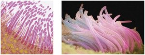
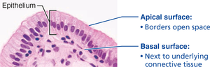
Polarity: Cells have distinct apical (top) and basal (bottom) surfaces. The apical surface may have microvilli, while the basal surface attaches to the basal lamina.
Specialized contacts: Cells are tightly joined by junctions (tight junctions, desmosomes).
Supported by connective tissue: The basement membrane (basal and reticular lamina) reinforces the sheet.
Avascular but innervated: No blood vessels, but supplied by nerves; nutrients diffuse from underlying tissues.
Regeneration: High capacity for renewal, especially in areas exposed to friction or hostile environments.
Classification of Epithelial Tissue
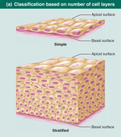
Epithelial tissues are classified by the number of cell layers and cell shape:
Simple epithelium: Single layer, ideal for absorption and filtration.
Stratified epithelium: Multiple layers, ideal for protection.
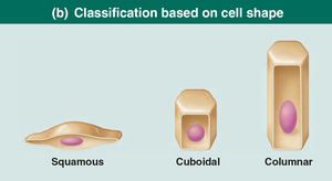
Cell shapes: Squamous (flat), cuboidal (cube-like), columnar (tall).
Types of Epithelial Tissue
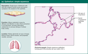
Simple Squamous Epithelium: Thin, flat cells for rapid diffusion; found in kidneys, lungs, blood vessels (endothelium), and serous membranes (mesothelium).
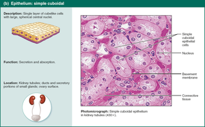
Simple Cuboidal Epithelium: Cube-shaped cells for secretion and absorption; found in kidney tubules and gland ducts.
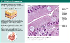
Simple Columnar Epithelium: Tall cells, often with microvilli or cilia; involved in absorption and secretion; found in digestive tract, gallbladder, bronchi, and uterine tubes.
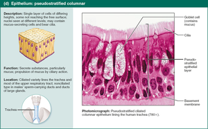
Pseudostratified Columnar Epithelium: Appears multilayered but is single-layered; often ciliated; found in respiratory tract and large gland ducts.
Stratified Squamous Epithelium: Multiple layers; protects against abrasion; found in skin (keratinized) and moist linings (nonkeratinized).
Stratified Cuboidal/Columnar Epithelium: Rare; found in sweat glands, mammary glands, pharynx, male urethra.
Transitional Epithelium: Changes shape when stretched; lines urinary organs (bladder, ureters, urethra).
Glandular Epithelia
Types of Glands
A gland is one or more cells that secrete an aqueous fluid. Glands are classified by site of product release (endocrine or exocrine) and number of cells (unicellular or multicellular).
Endocrine glands: Ductless, release hormones into interstitial fluid and blood.
Exocrine glands: Release products onto surfaces or into cavities via ducts; can be unicellular (goblet cells) or multicellular (salivary glands).
Connective Tissue
Overview and Functions
Connective tissue is the most abundant tissue, providing support, protection, insulation, storage, and transport. Four main classes: connective tissue proper, cartilage, bone, and blood.
Connective tissue proper: Loose (areolar, adipose, reticular) and dense (regular, irregular, elastic).
Cartilage: Hyaline, elastic, fibrocartilage.
Bone: Osseous tissue.
Blood: Fluid tissue for transport.
Common Characteristics
Extracellular matrix: Nonliving material supporting cells.
Common origin: All arise from mesenchyme.
Vascularity: Varies from avascular (cartilage) to highly vascular (bone).
Structural Elements
Ground substance: Gel-like material with interstitial fluid, cell adhesion proteins, and proteoglycans.
Fibers: Collagen (strength), elastic (stretch), reticular (support).
Cells: Immature "-blast" cells (secrete matrix), mature "-cyte" cells (maintain matrix), adipocytes, leukocytes, mast cells, macrophages.
Muscle Tissue
Types and Functions
Muscle tissue is highly vascularized and responsible for movement. Muscle cells contain myofilaments (actin and myosin) for contraction. Three types:
Skeletal muscle: Voluntary, striated, multinucleated; moves bones.
Cardiac muscle: Involuntary, striated, one nucleus, intercalated discs; found in heart.
Smooth muscle: Involuntary, non-striated, spindle-shaped; found in walls of hollow organs.
Nervous Tissue
Structure and Function
Nervous tissue is the main component of the nervous system, regulating and controlling body functions. It consists of neurons (transmit electrical signals) and glial cells (support, insulate, protect neurons).
Membranes
Types of Membranes
Membranes are organs composed of more than one tissue type, covering and lining body surfaces. Three main types:
Cutaneous membrane: Skin; dry, keratinized stratified squamous epithelium attached to connective tissue.
Mucous membrane: Lines cavities open to exterior; moist, often secretes mucus.
Serous membrane: Lines closed ventral cavities; moist, simple squamous epithelium (mesothelium) on areolar connective tissue.
Tissue Repair
Repair Mechanisms
Tissue repair occurs via regeneration (restores original function) or fibrosis (scar tissue replaces original tissue, function lost). Steps include inflammation, organization (restores blood supply), and permanent repair.
Developmental Aspects of Tissues
Embryonic Germ Layers
Primary germ layers (ectoderm, mesoderm, endoderm) form early in development and specialize into the four primary tissues. Nerve tissue arises from ectoderm, muscle and connective tissue from mesoderm, and epithelial tissue from all three.
Summary Table: Epithelial Tissue Classification
Type | Structure | Function | Location |
|---|---|---|---|
Simple Squamous | Single layer, flat cells | Diffusion, filtration | Kidney, lungs, blood vessels |
Simple Cuboidal | Single layer, cube-shaped cells | Secretion, absorption | Kidney tubules, glands |
Simple Columnar | Single layer, tall cells | Absorption, secretion | Digestive tract, bronchi |
Pseudostratified Columnar | Single layer, varying heights | Secretion, movement of mucus | Respiratory tract |
Stratified Squamous | Multiple layers, flat cells | Protection | Skin, mouth, esophagus |
Transitional | Multiple layers, shape changes | Stretching | Bladder, ureters |
