 Back
BackChapter 4: Tissue – The Living Fabric (ANP College Study Notes)
Study Guide - Smart Notes
 Tailored notes based on your materials, expanded with key definitions, examples, and context.
Tissue: The Living Fabric
Introduction to Tissues
Tissues are groups of cells that are similar in structure and perform a common or related function. Understanding tissue types is essential for monitoring potential tissue damage and for clinical applications such as wound healing and disease diagnosis. The principle of structure and function complementarity is crucial in tissue biology.
The Four Basic Tissue Types
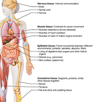
Epithelial Tissue: Covers surfaces, lines cavities, and forms glands.
Connective Tissue: Supports, binds, protects, and insulates other tissues.
Muscle Tissue: Produces movement.
Nervous Tissue: Controls and communicates information.
Epithelial Tissue
Definition and Functions
Epithelial tissue (epithelium) is a sheet of cells that covers body surfaces and lines cavities. It exists in two main forms: covering & lining epithelia (on external and internal surfaces) and glandular epithelia (secretory tissue in glands). Its main functions include protection, secretion, absorption, filtration, excretion, and sensory reception.
Special Characteristics of Epithelia
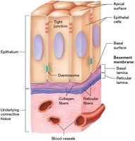
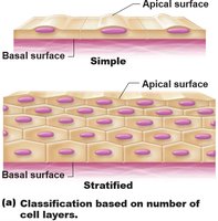
Polarity: Cells have distinct apical (upper) and basal (lower) surfaces, which differ in structure and function. The apical surface may have microvilli or cilia.
Supported by Connective Tissue: The basal surface is attached to a basement membrane, which reinforces the epithelial sheet and defines its boundary.
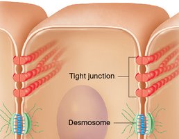
Specialized Contacts: Cells are tightly joined by junctions such as desmosomes and tight junctions, forming continuous sheets.
Avascular but Innervated: Epithelia lack blood vessels but are supplied by nerves.
High Regeneration Rate: Epithelial cells rapidly replace lost or damaged cells.
Classification of Epithelia
Epithelia are classified by two criteria: the number of cell layers and the shape of the cells in the apical layer.
Number of Cell Layers:
Simple epithelia: One layer of cells.
Stratified epithelia: Two or more layers of cells.
Cell Shape:
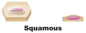
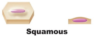
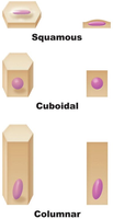
Squamous: Flattened, scale-like cells.
Cuboidal: Cube-shaped cells with round, central nuclei.
Columnar: Taller than wide, column-shaped cells with elongated nuclei.
Types of Epithelial Tissue
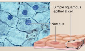
Simple Squamous Epithelium: Single layer of flattened cells; suited for diffusion and filtration. Found in air sacs of lungs, lining of blood vessels (endothelium), and serous membranes (mesothelium).

Simple Cuboidal Epithelium: Single layer of cube-shaped cells; functions in absorption and secretion. Found in kidney tubules and ducts of glands.

Simple Columnar Epithelium: Single layer of tall cells; functions in absorption and secretion. Nonciliated type lines most of the digestive tract; ciliated type lines small bronchi and uterine tubes.
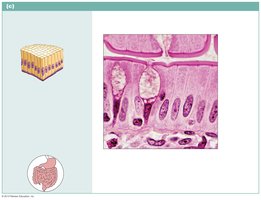
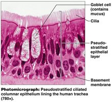
Pseudostratified Columnar Epithelium: Appears stratified but all cells rest on the basement membrane; functions in secretion and propulsion of mucus. Found in trachea and upper respiratory tract.
Stratified Squamous Epithelium: Multiple layers; protects against abrasion. Keratinized type forms the skin; nonkeratinized type lines the mouth, esophagus, and vagina.
Transitional Epithelium: Specialized for stretching; lines the urinary bladder, ureters, and part of the urethra.
Glandular Epithelia
Glands are specialized cells or organs of epithelial origin that secrete products (secretions). Glands are classified by where they release their product and by the number of cells.
Exocrine Glands: Have ducts; secrete products locally (e.g., sweat, oil, mucus, salivary glands).
Endocrine Glands: Ductless; secrete hormones into interstitial fluid and blood for systemic effects.
Unicellular Exocrine Glands: Goblet and mucous cells; produce mucin for lubrication.
Multicellular Exocrine Glands: Classified by duct structure (simple or compound) and secretory unit shape (tubular, alveolar, tubuloalveolar).
Modes of Secretion:
Merocrine: Secrete by exocytosis (e.g., salivary glands).
Holocrine: Accumulate products until cell ruptures (e.g., sebaceous glands).
Connective Tissue
Definition and Functions
Connective tissue is the most abundant and widely distributed tissue in the body. It supports, binds, protects, insulates, stores reserve fuel, and transports substances. It develops from embryonic mesenchyme.
Characteristics of Connective Tissue
Degrees of Vascularity: Varies from avascular (cartilage) to highly vascular (bone).
Extracellular Matrix (ECM): Non-living material consisting of ground substance (gel-like), fibers (collagen, elastic, reticular), and cells.
Cell Types: Immature "-blast" cells (e.g., fibroblasts, chondroblasts, osteoblasts) secrete ECM; mature "-cyte" cells maintain matrix.
Connective Tissue Fibers
Collagen: Strongest and most abundant; provides high tensile strength.
Elastic: Networks of elastin fibers; allow stretch and recoil.
Reticular: Short, fine collagenous fibers; connect connective tissue to adjacent tissues.
Major Classes of Connective Tissue
Connective Tissue Proper: Loose (areolar, adipose, reticular) and dense (regular, irregular, elastic).
Cartilage: Hyaline, elastic, fibrocartilage; avascular, receives nutrients from perichondrium.
Bone (Osseous Tissue): Compact and spongy; supports and protects.
Blood: Fluid connective tissue; transports gases, nutrients, wastes.
Muscle Tissue
Types and Functions
Muscle tissue is specialized for contraction and movement. There are three types:
Skeletal Muscle: Voluntary control; attached to bones; striated.
Cardiac Muscle: Involuntary control; found in the heart; striated; contains intercalated discs with desmosomes and gap junctions.
Smooth Muscle: Involuntary control; found in walls of hollow organs; non-striated.
Nervous Tissue
Structure and Function
Nervous tissue consists of neurons and neuroglia. Neurons transmit electrical impulses, while neuroglia support, insulate, and protect neurons. Nervous tissue is found in the brain, spinal cord, and nerves.
Epithelial Membranes
Types of Membranes
Epithelial membranes are simple organs composed of an epithelium bound to underlying connective tissue. There are three types:
Cutaneous Membrane: Dry; skin; consists of keratinized stratified squamous epithelium and dense irregular connective tissue.
Mucous Membrane: Wet; lines body cavities open to the exterior; functions in protection, secretion, and absorption.
Serous Membrane: Wet; lines closed ventral body cavities; secretes serous fluid for lubrication.
Tissue Repair
Steps in Tissue Repair
Tissue repair occurs via regeneration (replacement by same tissue) or fibrosis (replacement by scar tissue). The process involves three steps:
Inflammation: Sets the stage; release of chemicals causes swelling, redness, heat, and pain.
Organization: Restores blood supply; granulation tissue forms, fibroblasts produce collagen, debris is removed.
Regeneration and Fibrosis: Permanent repair; epithelium regenerates, scar tissue may remain.
Regenerative Capacity of Tissues
High Capacity: Epithelial tissues, bone, areolar connective tissue, dense irregular connective tissue, blood-forming tissue.
Moderate Capacity: Smooth muscle, dense regular connective tissue.
Weak Capacity: Skeletal muscle, cartilage.
No Functional Capacity: Cardiac muscle, nervous tissue of CNS (leads to fibrosis).
