 Back
Chapter 4: Tissue – The Living Fabric (Mini-Textbook Study Notes)
Study Guide - Smart Notes
 Tailored notes based on your materials, expanded with key definitions, examples, and context.
Tissue: The Living Fabric
Overview of Tissue Types
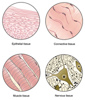
Tissues are groups of cells that are similar in structure and work together to perform specific functions. The human body contains four primary tissue types, each with distinct roles:
Epithelial tissue: Covers and lines surfaces and cavities.
Connective tissue: Supports, binds, and protects other tissues.
Muscle tissue: Contracts to produce movement.
Nervous tissue: Communicates signals to coordinate body functions.
Characteristics of Tissues
Each tissue type has unique structural and functional characteristics. For example, epithelial tissue forms protective boundaries, connective tissue provides structural support, muscle tissue enables movement, and nervous tissue transmits electrical impulses.
Epithelial Tissue
General Features
Epithelial tissue (epithelium; plural epithelia) is a thin, continuous, protective layer of cells. It forms boundaries between different environments and is classified into two main forms:
Covering and lining epithelia: Lines surfaces and cavities throughout the body.
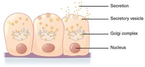
Glandular epithelia: Specialized for secretion, forming glands.
Classification of Epithelial Tissue
Epithelial tissues are classified based on cell shape and the number of cell layers:
Cell shapes: Squamous (flat), cuboidal (cube-shaped), columnar (tall).
Number of layers: Simple (single layer) or stratified (multiple layers).
Examples of epithelial tissue types:
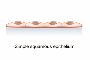
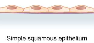
Simple squamous epithelium: Single layer of flat cells.
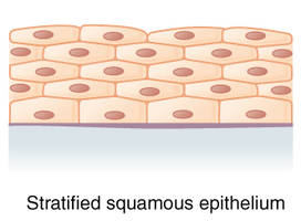
Stratified squamous epithelium: Multiple layers of flat cells.
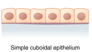
Simple cuboidal epithelium: Single layer of cube-shaped cells.
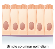
Simple columnar epithelium: Single layer of tall, column-like cells.
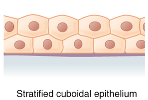
Stratified cuboidal epithelium: Multiple layers of cube-shaped cells.
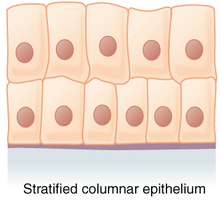
Stratified columnar epithelium: Multiple layers of column-like cells.
Structural Features of Epithelia
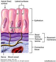
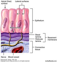
Epithelial tissues exhibit apical-basal polarity, meaning they have an apical surface (facing the lumen or outside) and a basal surface (attached to underlying connective tissue). The basement membrane separates the epithelium from connective tissue and consists of the basal lamina (next to epithelium) and reticular lamina (next to connective tissue).
Other Characteristics of Epithelia
Avascular but innervated: Epithelial tissues lack blood vessels but are supplied by nerves. They rely on nourishment from underlying connective tissue.
Regenerative: Epithelial cells rapidly replace lost cells by cell division.
Specialized contacts: Cells are held together by tight junctions and desmosomes.
Simple Squamous Epithelium
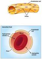
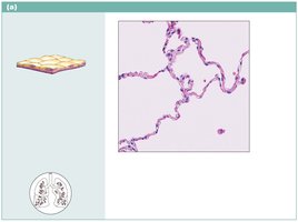
Simple squamous epithelium consists of a single thin layer of flat cells, designed for rapid exchange of substances via diffusion and filtration. It is found in locations where quick transport is necessary, such as air sacs of lungs, inner lining of blood vessels, and serous membranes.
Mesothelium: Lines serous membranes of ventral body cavities.
Endothelium: Lines lymphatic vessels, blood vessels, and heart.
Glandular Epithelia
General Features
A gland is a single epithelial cell or group of epithelial cells adapted for secretion. Glands are classified by:
Site of product release: Endocrine (ductless) or exocrine (with ducts).
Number of cells: Unicellular or multicellular.
Endocrine Glands
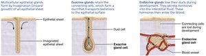
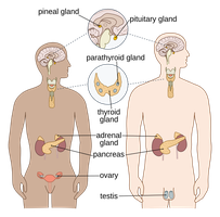
Endocrine glands are ductless glands that secrete hormones directly into the interstitial fluid, which are then taken up into the bloodstream. Hormones act as messenger chemicals, traveling through the circulatory system to reach target organs.
Exocrine Glands
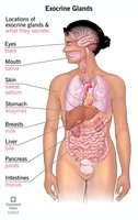
Exocrine glands secrete products onto body surfaces or into body cavities via ducts. Examples include mucous, sweat, oil, and salivary glands.
Unicellular Exocrine Glands
Unicellular exocrine glands, such as mucous cells and goblet cells, are found in epithelial linings of intestinal and respiratory tracts. They produce mucin, a glycosylated protein that forms mucus.
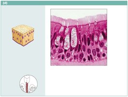
Multicellular Exocrine Glands
Multicellular exocrine glands are composed of a duct and a secretory unit. They are classified by structure (tubular or alveolar/acinar) and mode of secretion (merocrine or holocrine).
Tubular: Tube-like secretory unit.
Alveolar (acinar): Sac or pouch-shaped secretory unit.
They are also classified by duct branching:
Simple: Unbranched duct.
Compound: Branching duct.
Modes of Secretion
Merocrine glands: Products are secreted by exocytosis; cells remain intact. Examples: sweat glands, salivary glands, pancreatic exocrine glands.
Holocrine glands: Products are secreted by rupture of gland cells; cells are destroyed. Example: sebaceous (oil) glands.
Connective Tissue
General Features
Connective tissue (CT) is the most abundant and widely distributed tissue type in the body. It serves functions such as binding and support, protection, insulation, energy storage (fat), and transport of substances (blood).
Classification of Connective Tissue
Connective tissue proper: Includes areolar, adipose, fibrous CT, etc.
Cartilage: Specialized for support and flexibility (see Ch. 6).
Bone tissue: Provides structural support (see Ch. 6).
Blood: Specialized for transport (see A&P II).
Characteristics of Connective Tissue
Originates from mesenchyme (embryonic CT).
Exhibits varying degrees of vascularity (from avascular cartilage to highly vascularized bone).
Cells are separated by an extracellular matrix composed of ground substance and protein fibers.
Extracellular Matrix
The extracellular matrix consists of:
Ground substance: Gel-like, nonfibrous part containing interstitial fluid, adhesion glycoproteins, and proteoglycans (affect viscosity).
Protein fibers: Collagen (strength), elastic (stretch/recoil), reticular (support).
Connective Tissue Proper
Connective tissue proper is divided into two subclasses:
Loose connective tissues: Areolar, adipose, reticular.
Dense (fibrous) connective tissues: Dense regular, dense irregular, elastic.
Dense Regular Connective Tissue
Dense regular connective tissue consists of parallel collagen fibers and a few elastic fibers, with fibroblasts as the main cell type. It withstands high tension and stretching and is found in tendons and ligaments.
Dense Irregular Connective Tissue
Dense irregular connective tissue has randomly arranged collagen fibers and a few elastic fibers, with fibroblasts. It resists tension from many directions and is found in the dermis of skin, joint capsules, and fibrous coverings of some organs.
Body Membranes
Overview
Body membranes are thin layers of tissue that line body surfaces and cavities. They are classified into epithelial and connective membranes. Epithelial membranes are composed of both epithelial and connective tissue types.
Cutaneous membrane: Skin; a dry membrane.
Mucous membranes: Line body tracts open to the exterior (digestive, respiratory, urogenital).
Serous membranes: Line closed body cavities (thoracic, abdominal).
Cutaneous Membrane (Skin)
The cutaneous membrane consists of an epithelial layer (epidermis: keratinized stratified squamous epithelium) and a connective tissue layer (dermis).
Mucous Membranes
Mucous membranes line body tracts open to the exterior. They consist of a mucosal epithelium (cell type and layers vary) and a connective tissue layer (lamina propria: loose areolar CT).
Serous Membranes
Serous membranes line closed body cavities. They consist of a mesothelium (simple squamous epithelium) and a submesothelial connective tissue layer (loose areolar CT). Examples include the pleurae (lungs), pericardium (heart), and peritoneum (abdominopelvic organs).
Tissue Repair
Regeneration and Fibrosis
Tissue repair occurs via two major mechanisms:
Regeneration: Destroyed tissue is replaced with the same kind of tissue; original function is restored.
Fibrosis: Destroyed tissue is replaced with scar tissue; original function is lost.
Regenerative Capacity of Tissues
High regenerative capacity: Epithelial tissues, bone, areolar CT, dense irregular CT, blood-forming tissue.
No functional regenerative capacity: Cardiac muscle and nervous tissue.
Steps of Tissue Repair (Skin)
Inflammation: Damaged tissues release inflammatory chemicals, causing dilation of blood vessels, increased vessel permeability, redness, heat, swelling, pain, and blood clotting.
Proliferation (organization): Formation of granulation tissue (highly vascular connective tissue), production of collagen fibers, proliferation of epithelial cells, and phagocytosis of debris.
Remodeling: Regeneration replaces destroyed tissue with the same type; fibrosis replaces it with scar tissue.
Additional info: These notes provide a comprehensive overview of tissue types, their classification, structural features, and repair mechanisms, suitable for exam preparation in an anatomy and physiology college course.
