 Back
Chapter 4: Tissue – The Living Fabric (Epithelial Tissue)
Study Guide - Smart Notes
 Tailored notes based on your materials, expanded with key definitions, examples, and context.
Tissue: The Living Fabric
Introduction to Tissues
Tissues are groups of cells with similar structure and function, essential for maintaining homeostasis in the human body. Understanding tissue types is crucial for monitoring health and diagnosing conditions such as bedsores.
Definition: Tissues are collections of cells that perform common or related functions.
Histology: The study of tissues.
Four Basic Tissue Types: Epithelial, connective, muscle, and nervous tissue.
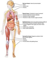
Microscopy of Human Tissue
Microscopy is essential for studying tissues, requiring preparation steps to ensure visibility and contrast.
Fixation: Preserves tissue with solvents.
Sectioning: Slices tissue thin enough for light or electron transmission.
Staining: Enhances contrast; light microscopy uses colored dyes, electron microscopy uses heavy metals.
Artifacts: Distortions introduced during preparation.
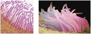
Epithelial Tissue
Definition and Functions
Epithelial tissue forms boundaries between different environments, covering surfaces and lining cavities. It is specialized for protection, absorption, filtration, excretion, secretion, and sensory reception.
Covering and Lining Epithelia: Found on external and internal surfaces (e.g., skin).
Glandular Epithelia: Secretory tissue in glands (e.g., salivary glands).
Special Characteristics of Epithelial Tissues
Epithelial tissues possess five key characteristics that distinguish them from other tissue types.
Polarity: Cells have distinct apical (top) and basal (bottom) surfaces. The apical surface may have microvilli, increasing surface area for absorption. The basal surface attaches to the basal lamina, anchoring the tissue.
Specialized Contacts: Cells are tightly joined by junctions (tight junctions and desmosomes) to form continuous sheets.
Supported by Connective Tissues: The basement membrane (basal and reticular lamina) reinforces the epithelial sheet and defines boundaries.
Avascular but Innervated: Epithelial tissues lack blood vessels but are supplied by nerves. Nutrients diffuse from underlying connective tissue.
Regeneration: High regenerative capacity, especially in areas exposed to friction or hostile environments.
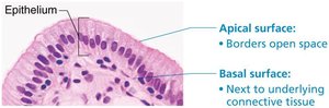
Classification of Epithelia
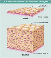
Epithelial tissues are classified by the number of cell layers and cell shape.
Number of Layers:
Simple Epithelium: Single layer, specialized for absorption, secretion, and filtration.
Stratified Epithelium: Multiple layers, specialized for protection.
Cell Shape:
Squamous: Flattened, scale-like.
Cuboidal: Box-like, cube-shaped.
Columnar: Tall, column-shaped.
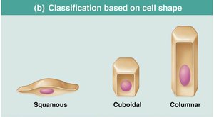
Types of Epithelial Tissue
Simple Epithelia
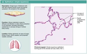
Simple Squamous Epithelium: Single layer of flattened cells, allows rapid diffusion and filtration. Locations: Kidney glomeruli, air sacs of lungs, lining of heart, blood vessels, lymphatic vessels. Special Types:
Endothelium: Lines blood vessels and heart.
Mesothelium: Lines serous membranes in ventral body cavity.
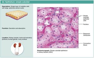
Simple Cuboidal Epithelium: Single layer of cube-shaped cells, functions in secretion and absorption. Locations: Kidney tubules, ducts of glands.
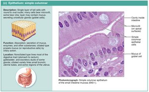
Simple Columnar Epithelium: Single layer of tall cells, often with microvilli or cilia, and goblet cells. Functions in absorption and secretion. Locations: Digestive tract, gallbladder, ducts of glands, bronchi, uterine tubes.
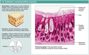
Pseudostratified Columnar Epithelium: Appears multi-layered but is actually a single layer. Often ciliated, contains goblet cells. Functions in secretion and movement of mucus. Locations: Upper respiratory tract, ducts of large glands, tubules in testes.
Stratified Epithelia
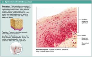
Stratified Squamous Epithelium: Multiple layers, surface cells are squamous. Protects against abrasion. Locations: Skin (keratinized), moist linings of esophagus, mouth, vagina (nonkeratinized).
Stratified Cuboidal and Columnar Epithelium: Rare, found in sweat glands, mammary glands, pharynx, male urethra, and glandular ducts.
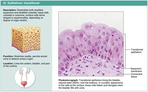
Transitional Epithelium: Resembles both stratified squamous and cuboidal. Stretches readily, permits stored urine to distend urinary organ. Locations: Lines ureters, bladder, and part of urethra.
Glandular Epithelia
Definition and Classification
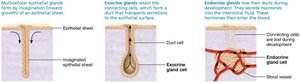
A gland is one or more cells that makes and secretes an aqueous fluid called a secretion. Glands are classified by site of product release (endocrine or exocrine) and number of cells (unicellular or multicellular).
Endocrine Glands: Ductless, release hormones into interstitial fluid, which are picked up by blood or lymph.
Exocrine Glands: Release products onto body surfaces or into cavities via ducts. Includes mucous, sweat, oil, and salivary glands.
Unicellular Exocrine Glands
The only important unicellular exocrine glands are mucous cells and goblet cells, which produce mucin that forms mucus.
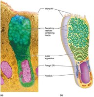
Multicellular Exocrine Glands
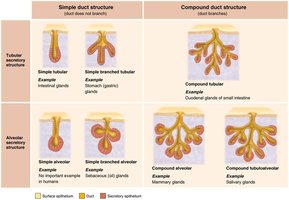
Multicellular exocrine glands consist of a duct and a secretory unit, often surrounded by connective tissue. They are classified by structure and mode of secretion.
Structure:
Simple glands: Unbranched ducts.
Compound glands: Branched ducts.
Tubular: Secretory cells form tubes.
Alveolar: Secretory cells form sacs.
Tubuloalveolar: Both types present.
Mode of Secretion:
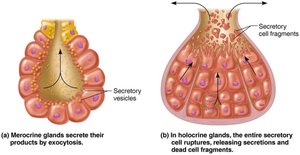
Merocrine: Secrete by exocytosis (e.g., sweat, pancreas).
Holocrine: Accumulate products and rupture (e.g., sebaceous glands).
Apocrine: Apex ruptures; existence in humans is controversial (possibly mammary glands).
Clinical Relevance
Homeostatic Imbalance: Cancer
Cancerous epithelial cells can penetrate the basement membrane and invade underlying tissues, leading to metastasis and spread of cancer.
Summary Table: Epithelial Tissue Types
Type | Structure | Function | Location |
|---|---|---|---|
Simple Squamous | Single layer, flat cells | Diffusion, filtration | Lungs, blood vessels |
Simple Cuboidal | Single layer, cube cells | Secretion, absorption | Kidney tubules, glands |
Simple Columnar | Single layer, tall cells | Absorption, secretion | Digestive tract |
Pseudostratified Columnar | Single layer, varying heights | Mucus secretion, movement | Respiratory tract |
Stratified Squamous | Multiple layers, flat surface cells | Protection | Skin, esophagus |
Transitional | Multiple layers, shape changes | Stretching | Bladder, ureters |
Key Terms and Concepts
Basement Membrane: Layer anchoring epithelium to connective tissue.
Microvilli: Fingerlike projections increasing surface area.
Goblet Cells: Unicellular glands producing mucus.
Merocrine, Holocrine, Apocrine: Modes of glandular secretion.
Equations and Formulas
While epithelial tissue does not involve mathematical equations, the following formula is relevant for diffusion across simple squamous epithelium:
Where: J = flux (rate of diffusion), D = diffusion coefficient, C = concentration, x = distance.
Conclusion
Epithelial tissue is fundamental to the structure and function of the human body, providing protection, absorption, secretion, and more. Understanding its classification, characteristics, and clinical relevance is essential for students of anatomy and physiology.
