 Back
BackChapter 4: Tissue – The Living Fabric (Epithelial Tissue)
Study Guide - Smart Notes
 Tailored notes based on your materials, expanded with key definitions, examples, and context.
Tissue: The Living Fabric
Introduction to Tissues
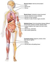
Tissues are groups of cells with similar structure and function, essential for maintaining homeostasis in the human body. The study of tissues is known as histology. There are four basic tissue types: epithelial, connective, muscle, and nervous tissue.
Epithelial tissue: Covers surfaces and lines cavities.
Connective tissue: Supports and binds other tissues.
Muscle tissue: Facilitates movement.
Nervous tissue: Enables communication and control.
Microscopy of Human Tissue
To study tissues, samples must be prepared for microscopy:
Fixation: Preserves tissue structure.
Sectioning: Slices tissue thinly for light or electron transmission.
Staining: Enhances contrast; light microscopy uses dyes, electron microscopy uses heavy metals.
Epithelial Tissue
Definition and Functions
Epithelial tissue (epithelium) is a sheet of cells covering body surfaces or lining cavities. It exists in two main forms:
Covering and lining epithelia: External and internal surfaces (e.g., skin).
Glandular epithelia: Secretory tissue in glands (e.g., salivary glands).
Main functions include protection, absorption, filtration, excretion, secretion, and sensory reception.
Special Characteristics of Epithelial Tissues
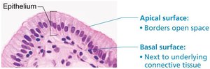
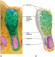
Polarity: Cells have distinct apical (top) and basal (bottom) surfaces. The apical surface may have microvilli, increasing surface area for absorption. The basal surface attaches to the basal lamina, anchoring the tissue.
Specialized contacts: Cells are tightly joined by junctions (tight junctions, desmosomes) to form continuous sheets.
Supported by connective tissues: The basement membrane (basal and reticular lamina) reinforces the epithelium and defines boundaries.
Avascular but innervated: Epithelial tissues lack blood vessels but are supplied by nerves; nutrients diffuse from underlying connective tissue.
Regeneration: High capacity for renewal, especially in areas exposed to friction or hostile environments.
Classification of Epithelia
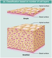
Epithelial tissues are classified by cell layers and cell shape:
Number of cell layers:
Simple epithelium: Single layer, specialized for absorption, secretion, filtration.
Stratified epithelium: Multiple layers, specialized for protection.
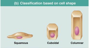
Cell shape:
Squamous: Flattened, scale-like.
Cuboidal: Cube-shaped.
Columnar: Tall, column-like.
Types of Epithelial Tissue
Simple Epithelia
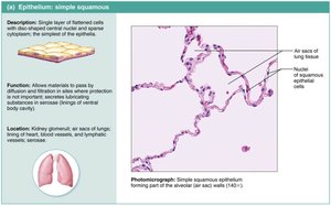
Simple squamous epithelium: Single layer of flat cells; allows rapid diffusion and filtration. Found in kidneys, lungs, blood vessels (endothelium), and serous membranes (mesothelium).
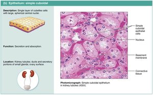
Simple cuboidal epithelium: Single layer of cube-shaped cells; functions in secretion and absorption. Found in kidney tubules and gland ducts.
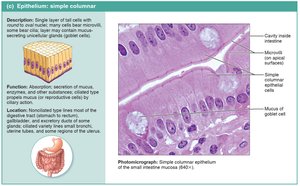
Simple columnar epithelium: Single layer of tall cells; may have microvilli or cilia, and goblet cells. Functions in absorption and secretion. Found in digestive tract, gallbladder, bronchi, uterine tubes.
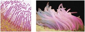
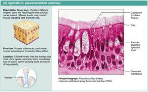
Pseudostratified columnar epithelium: Appears multi-layered but is a single layer; often ciliated and contains goblet cells. Functions in secretion and movement of mucus. Found in upper respiratory tract, large gland ducts, and tubules in testes.
Stratified Epithelia
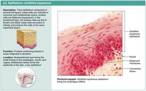
Stratified squamous epithelium: Multiple layers; protects against abrasion. Keratinized type found in skin, nonkeratinized in moist linings (e.g., esophagus).
Stratified cuboidal epithelium: Rare; found in sweat and mammary glands.
Stratified columnar epithelium: Rare; found in pharynx, male urethra, and some glandular ducts.
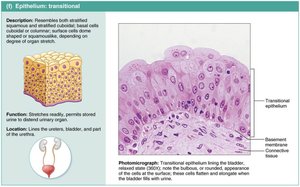
Transitional epithelium: Resembles both stratified squamous and cuboidal; stretches readily, found in urinary organs (bladder, ureters, urethra).
Glandular Epithelia
Definition and Classification
A gland is one or more cells that secrete an aqueous fluid called a secretion. Glands are classified by:
Site of product release:
Endocrine: Internally secreting (hormones).
Exocrine: Externally secreting (sweat, oil).
Number of cells:
Unicellular: Goblet cells.
Multicellular: Salivary glands.
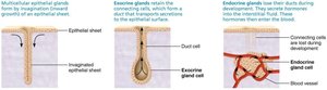
Endocrine Glands
Ductless; secrete hormones into interstitial fluid, which is absorbed by blood or lymph.
Hormones act on specific target organs.
Exocrine Glands
Secrete products onto body surfaces or into cavities via ducts.
Include mucous, sweat, oil, and salivary glands.
Can be unicellular or multicellular.
Unicellular Exocrine Glands
Goblet cells and mucous cells are the main unicellular exocrine glands.
Produce mucin, which forms mucus for protection and lubrication.
Multicellular Exocrine Glands
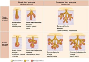
Composed of a duct and secretory unit, often surrounded by connective tissue.
Classified by structure (simple or compound ducts; tubular, alveolar, or tubuloalveolar) and mode of secretion.
Modes of Secretion
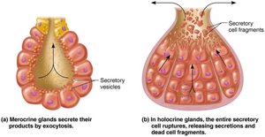
Merocrine: Secrete by exocytosis (e.g., sweat, pancreas).
Holocrine: Accumulate products and rupture (e.g., sebaceous glands).
Apocrine: Apex ruptures; existence in humans is debated (possibly mammary glands).
Summary Table: Epithelial Tissue Types
Type | Structure | Function | Location |
|---|---|---|---|
Simple squamous | Single layer, flat cells | Diffusion, filtration | Kidney, lungs, blood vessels |
Simple cuboidal | Single layer, cube cells | Secretion, absorption | Kidney tubules, glands |
Simple columnar | Single layer, tall cells | Absorption, secretion | Digestive tract, gallbladder |
Pseudostratified columnar | Single layer, varying heights | Mucus secretion, movement | Respiratory tract |
Stratified squamous | Multiple layers, flat cells | Protection | Skin, esophagus |
Transitional | Multiple layers, variable shape | Stretching | Bladder, ureters |
Clinical Relevance
Cancerous epithelial cells can breach the basement membrane, invading underlying tissues and spreading cancer. Monitoring tissue health is crucial in clinical practice, such as preventing bedsores.
Key Terms and Concepts
Histology: Study of tissues.
Basement membrane: Structure supporting epithelial tissue.
Microvilli: Fingerlike projections increasing surface area.
Goblet cell: Unicellular gland producing mucus.
Merocrine, holocrine, apocrine: Modes of glandular secretion.
Equations and Scientific Notation
No specific equations are required for this chapter, but cell division and diffusion principles are relevant:
Diffusion rate: where is diffusion rate, is diffusion constant, is area, is concentration difference, is distance.
Additional info:
Some rare epithelial types (stratified cuboidal and columnar) are not illustrated but are briefly described. Apocrine secretion is controversial in humans.
