 Back
BackChapter 4: Tissue: The Living Fabric – Study Notes
Study Guide - Smart Notes
 Tailored notes based on your materials, expanded with key definitions, examples, and context.
Chapter 4: Tissue – The Living Fabric
Introduction to Tissues
Tissues are groups of cells similar in structure that perform common or related functions, maintaining homeostasis in the body. The study of tissues is called histology. There are four basic tissue types: epithelial, connective, muscle, and nervous tissue.
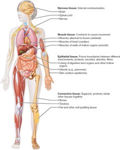
Epithelial Tissue
Definition and Main Categories
Epithelial tissue (epithelium) is a sheet of cells that covers body surfaces or lines body cavities. It exists in two main forms:
Covering and lining epithelium: Forms the outer layer of the skin and lines open cavities and organs.
Glandular epithelium: Forms glands of the body (e.g., salivary glands).
Main functions include protection, absorption, filtration, excretion, secretion, and sensory reception.
Special Characteristics of Epithelial Tissues
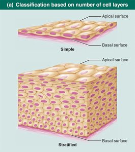
Polarity: Cells have an apical (top, exposed) and basal (bottom, attached) surface. The apical surface may have microvilli; the basal surface attaches to the basal lamina.
Specialized contacts: Cells fit closely together, forming continuous sheets with tight junctions and desmosomes.
Supported by connective tissue: All epithelial sheets rest on and are supported by connective tissue (basement membrane = basal lamina + reticular lamina).
Avascular but innervated: No blood vessels; nutrients diffuse from underlying tissues. Supplied by nerve fibers.
Regeneration: High regenerative capacity, especially in areas exposed to friction or hostile environments.

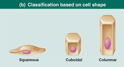
Classification of Epithelial Tissue
Epithelia are classified by the number of cell layers and cell shape:
Simple epithelia: Single cell layer (for absorption, secretion, filtration).
Stratified epithelia: Two or more layers (for protection in high-abrasion areas).
Cell shapes: Squamous (flat), cuboidal (cube-like), columnar (tall).
Types of Epithelia
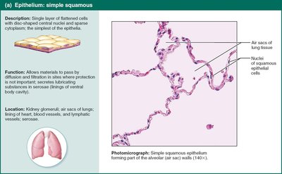
Simple Squamous Epithelium: Single layer of flat cells; allows rapid diffusion (e.g., alveoli of lungs, kidney glomeruli, endothelium, mesothelium).
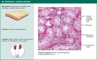
Simple Cuboidal Epithelium: Single layer of cube-shaped cells; secretion and absorption (e.g., kidney tubules, ducts of glands).
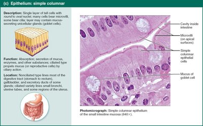
Simple Columnar Epithelium: Single layer of tall cells; absorption and secretion, may have microvilli or cilia (e.g., digestive tract, gallbladder, uterine tubes).
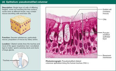
Pseudostratified Columnar Epithelium: Appears multilayered but is single-layered; often ciliated, secretes and moves mucus (e.g., upper respiratory tract).
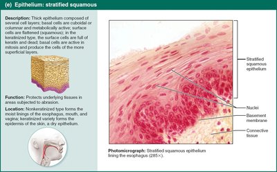
Stratified Squamous Epithelium: Multiple layers; protects underlying tissues (e.g., skin, mouth, esophagus).
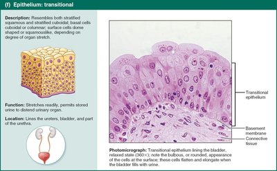
Transitional Epithelium: Resembles both stratified squamous and cuboidal; stretches (e.g., bladder, ureters).
Glandular Epithelia
A gland consists of one or more cells that make and secrete an aqueous fluid (secretion). Glands are classified by:
Site of product release: Endocrine (ductless, hormones into blood/lymph) vs. exocrine (onto body surfaces or into cavities via ducts).
Number of cells: Unicellular (e.g., goblet cells) vs. multicellular (e.g., salivary glands).
Unicellular exocrine glands: Mucous and goblet cells; secrete mucin (forms mucus).
Multicellular exocrine glands: Composed of duct and secretory unit; classified by structure (simple/compound, tubular/alveolar/tubuloalveolar) and mode of secretion (merocrine, holocrine, apocrine).
Connective Tissue
Overview and Functions
Connective tissue is the most abundant and widely distributed tissue type. Major functions include binding/support, protection, insulation, energy storage, and transport (blood). Four main classes:
Connective tissue proper
Cartilage
Bone
Blood
Comparison of Classes of Connective Tissues
Tissue Class | Subclasses | Cells | Matrix | General Features |
|---|---|---|---|---|
Connective Tissue Proper | Loose (areolar, adipose, reticular); Dense (regular, irregular, elastic) | Fibroblasts, fibrocytes, adipocytes, defense cells | Gel-like; collagen, reticular, elastic fibers | Binding, resisting tension, energy storage |
Cartilage | Hyaline, elastic, fibrocartilage | Chondroblasts, chondrocytes | Gel-like; collagen, elastic fibers | Cushioning, support, avascular |
Bone | Compact, spongy | Osteoblasts, osteocytes | Calcified; collagen fibers | Support, protection, blood cell formation |
Blood | — | RBCs, WBCs, platelets | Plasma (fluid) | Transport of gases, nutrients, wastes |
Common Characteristics of Connective Tissue
Extracellular matrix: Nonliving material (ground substance + fibers) separates living cells; allows tissue to bear weight, tension, and abuse.
Common origin: All arise from mesenchyme (embryonic tissue).
Structural Elements of Connective Tissue
Ground substance: Unstructured material filling space between cells; contains interstitial fluid, cell adhesion proteins, and proteoglycans (e.g., chondroitin sulfate, hyaluronic acid).
Fibers: Collagen (strongest), elastic (stretch/recoil), reticular (supportive networks).
Cells: "-blast" (immature, matrix-secreting) and "-cyte" (mature, maintain matrix) forms; also adipocytes, leukocytes, mast cells, macrophages.
Types of Connective Tissue Proper
Loose connective tissue: Areolar (packing, support), adipose (fat storage, insulation), reticular (support for blood cells in lymphoid organs).
Dense connective tissue: Regular (tendons, ligaments), irregular (dermis, organ capsules), elastic (arteries, vertebral ligaments).
Cartilage
Hyaline cartilage: Most abundant; supports, reinforces (ends of long bones, nose, trachea).
Elastic cartilage: More elastic fibers; maintains shape (ear, epiglottis).
Fibrocartilage: Strong, resists compression (intervertebral discs, knee).
Bone (Osseous Tissue)
Supports, protects, stores fat, synthesizes blood cells.
Osteoblasts produce matrix; osteocytes maintain it.
Highly vascularized; contains inorganic calcium salts.
Blood
Most atypical connective tissue (fluid).
Transports gases, nutrients, wastes, hormones.
Contains RBCs, WBCs, platelets in plasma.
Muscle Tissue
Overview
Muscle tissue is highly vascularized and responsible for movement. Muscle cells contain myofilaments (actin and myosin) for contraction. Three types:
Skeletal muscle: Voluntary, striated, multinucleate, attached to bones.
Cardiac muscle: Involuntary, striated, one nucleus, intercalated discs, found in heart.
Smooth muscle: Involuntary, non-striated, spindle-shaped, one nucleus, found in walls of hollow organs.
Nervous Tissue
Overview
Nervous tissue is the main component of the nervous system (brain, spinal cord, nerves). It regulates and controls body functions. Two main cell types:
Neurons: Respond to stimuli and transmit electrical signals via dendrites and axons.
Supporting cells (neuroglia): Support, insulate, and protect neurons.
Membranes
Types of Covering and Lining Membranes
Cutaneous membrane: Skin; keratinized stratified squamous epithelium attached to connective tissue (dry membrane).
Mucous membranes (mucosae): Line body cavities open to exterior (digestive, respiratory, urogenital tracts); moist, may secrete mucus.
Serous membranes (serosae): Line closed ventral body cavities; simple squamous epithelium (mesothelium) on areolar connective tissue; parietal (lines cavity) and visceral (covers organs) layers with serous fluid between.
Tissue Repair
Process of Tissue Repair
Regeneration: Replacement of destroyed tissue with the same kind; restores function.
Fibrosis: Connective tissue replaces destroyed tissue; function lost.
Inflammation: Inflammatory chemicals released, blood vessels dilate, clotting occurs.
Organization: Blood clot replaced by granulation tissue, epithelium regenerates, fibroblasts bridge gap, debris removed.
Regeneration and fibrosis: Scab detaches, tissue matures, scar tissue may remain.
Regenerative Capacity of Tissues
High: Epithelial, bone, areolar, dense irregular, blood-forming tissue.
Moderate: Smooth muscle, dense regular connective tissue.
Low/None: Cardiac muscle, nervous tissue (brain, spinal cord).
Developmental Aspects of Tissues
Primary germ layers: Ectoderm (nervous), mesoderm (muscle, connective), endoderm (epithelial from all three).
Tissues function well through youth; aging leads to thinning epithelia, less efficient repair, atrophy, increased cancer risk.
