 Back
BackChapter 4: Tissues – Structure, Function, and Classification
Study Guide - Smart Notes
 Tailored notes based on your materials, expanded with key definitions, examples, and context.
Tissues and Tissue Types
Overview of Tissue Types
Tissues are collections of specialized cells and cell products organized to perform specific functions. All organs are derived from four fundamental tissue types:
Epithelial Tissue: Maintains physical barriers and controls permeability.
Connective Tissue: Supports, connects, and protects other tissues.
Muscular Tissue: Composed of cells that contract to produce movement.
Nervous Tissue: Conveys information via electrical impulses.
Epithelial Tissue
Structure and Functions
Epithelial tissues separate two compartments and control permeability, providing physical protection. Some epithelia contain sensory nerves for sensation, and many form exocrine or endocrine glands for secretion.
Avascular: Epithelia lack blood vessels.
Regeneration: Stem cells allow continuous replacement.
Integrity: Maintained by intercellular connections.
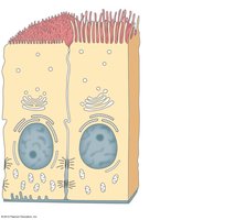
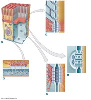
Intercellular Connections in Epithelia
Intercellular junctions are critical for epithelial integrity and function:
Tight Junctions: Form impermeable barriers.
Gap Junctions: Allow diffusion of ions and small molecules between cells.
Desmosomes: Tie adjacent cells together.
Hemidesmosomes: Attach cells to extracellular structures (basement membrane).
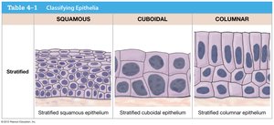
Morphological Classification of Epithelia
Epithelia are classified based on cell shape and number of layers:
Simple: One layer of cells.
Stratified: Multiple layers of cells.
Squamous: Flat cells.
Cuboidal: Cube-shaped cells.
Columnar: Tall, column-like cells.
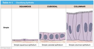
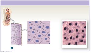
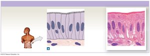
Simple Squamous Epithelia
Simple squamous epithelia are found in mesothelia lining body cavities, endothelia lining blood vessels, portions of kidney tubules, and alveoli of lungs. They reduce friction, control permeability, and facilitate absorption/secretion.
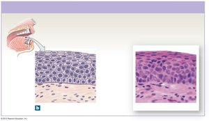
Stratified Squamous Epithelia
Located on the surface of skin and lining of mouth, throat, esophagus, rectum, anus, and vagina. Provides protection against abrasion, pathogens, and chemical attack.
Simple Cuboidal Epithelia
Found in portions of kidney tubules, ducts in exocrine glands, and endocrine glands. Functions include limited protection, secretion, and absorption.
Stratified Cuboidal Epithelia
Rare, found lining some gland ducts. Functions in protection, secretion, and absorption.
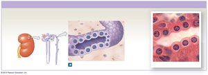
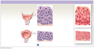
Transitional Epithelia
Located in the urinary bladder, renal pelvis, and ureters. Permits expansion and recoil after stretching.
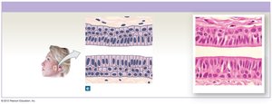
Simple Columnar Epithelia
Lines the stomach, intestine, and collecting ducts of kidneys. Functions in protection, secretion, and absorption.
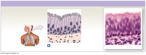
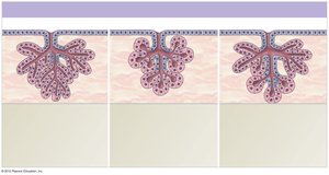
Pseudostratified Columnar Epithelia
Lines the nasal cavity, trachea, and bronchi. Functions in protection, secretion, and movement of mucus with cilia.
Stratified Columnar Epithelia
Found in small areas of the pharynx, epiglottis, and anus. Provides protection.
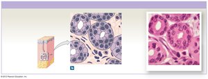
Glandular Epithelia
Glandular epithelia are specialized for secretion:
Endocrine Glands: Release hormones into interstitial fluid or blood; ductless.
Exocrine Glands: Secrete enzymes, mucins, or fluids onto epithelial surfaces via ducts.
Modes of Exocrine Secretion
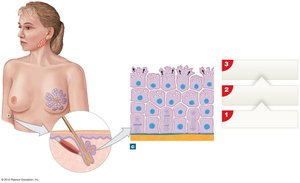
Merocrine: Secretion by exocytosis (e.g., salivary glands).
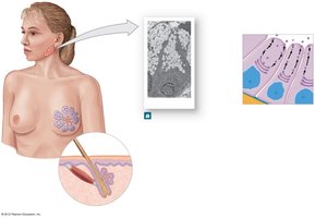
Apocrine: Secretion involves shedding of cytoplasm (e.g., mammary glands).
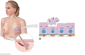
Holocrine: Secretion occurs as cells burst and are replaced by stem cells (e.g., sebaceous glands).

Types of Exocrine Secretion
Mucous Glands: Secrete mucins for lubrication.
Serous Glands: Produce watery, enzyme-rich secretions.
Mixed Glands: Produce both mucous and serous secretions.
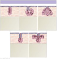
Structural Classification of Exocrine Glands
Exocrine glands are classified by their structure:
Simple Tubular: Intestinal glands
Simple Coiled Tubular: Merocrine sweat glands
Simple Branched Tubular: Gastric glands
Simple Branched Alveolar: Sebaceous glands
Compound Tubular: Seminiferous tubules in testes
Compound Alveolar/Acinar: Mammary glands
Compound Tubuloalveolar: Salivary glands, pancreas


Connective Tissue
Functions and Basic Components
Connective tissue connects different tissues, provides structural framework, protection, bulk transport, defense, and energy storage. Basic components include specialized cells, extracellular protein fibers, and ground substance (extracellular matrix).

Classification of Connective Tissues
Connective Tissue Proper: Loose and dense types; connects and protects.
Fluid Connective Tissues: Blood and lymph; transport.
Supporting Connective Tissues: Cartilage and bone; structural strength.
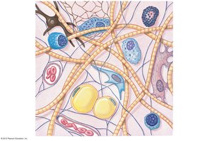
Cell Types in Connective Tissue Proper
Fibroblasts: Produce fibers and ground substance.
Fibrocytes: Inactive fibroblasts.
Adipocytes: Store lipids.
Mesenchymal Cells: Stem cells for regeneration.
Macrophages, Mast Cells, Lymphocytes: Immune functions.
Melanocytes: Store melanin pigment.
Fibers in Connective Tissue Proper
Collagen: Straight, unbranched, strong.
Reticular: Branched, meshwork.
Elastic: Branched, flexible.
Ground Substance: The matrix supporting cells, composed of proteoglycans, proteins, water, and minerals.
Connective Tissue Proper Structure
Loose Connective Tissue Proper
Areolar Tissue: Under skin, loosely packed, allows movement.
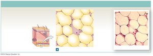
Adipose Tissue: Deep under skin, around organs; protection, insulation, energy storage.
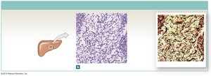
Reticular Tissue: Stroma of organs; supports parenchyma.


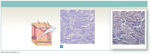
Dense Connective Tissue Proper
Dense Regular: Tendons, ligaments; transfers force.
Dense Irregular: Dermis, periosteum; resists multidirectional forces.
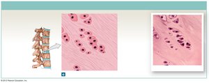
Elastic: Intervertebral ligaments, blood vessel walls; permits expansion/contraction.


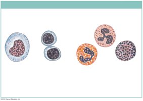
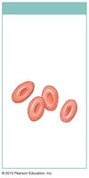
Fluid Connective Tissue: Blood and Lymph
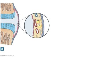
Blood
Blood contains formed elements (red and white blood cells, platelets) and plasma. Red blood cells (erythrocytes) transport oxygen, white blood cells (leukocytes) defend against infection, and platelets are important in clotting.
Lymph
Lymph is interstitial fluid entering lymphatic vessels to return to the cardiovascular system.
Supporting Connective Tissues: Cartilage and Bone
Cartilage
Cartilage matrix is a firm gel containing chondroitin sulfate and fibers. It is avascular, contains chondrocytes in lacunae, and is separated from surrounding tissues by perichondrium. Grows via interstitial and appositional growth.
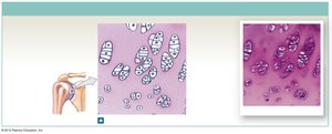
Hyaline Cartilage: Most articulations, respiratory tract; stiff, reduces friction.
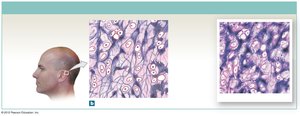
Elastic Cartilage: Auricle of external ear; flexible due to elastic fibers.
Fibrocartilage: Meniscus, intervertebral discs; resists compression.
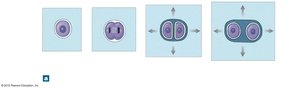
Growth of Cartilage
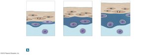
Interstitial Growth: Chondrocytes divide within lacunae, secrete matrix, and move apart.
Appositional Growth: New chondroblasts added from perichondrium, secrete matrix, become chondrocytes.

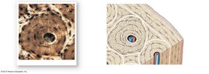
Bone
Bones contain osteocytes in lacunae, depend on canaliculi for nutrient diffusion, and have a dense mineralized matrix (mostly calcium phosphate). Surrounded by periosteum, bones have good blood supply and extensive remodeling and repair.
Comparison of Cartilage and Bone
Characteristic | Cartilage | Bone |
|---|---|---|
Cells | Chondrocytes | Osteocytes |
Ground Substance | Chondroitin sulfate, water | Calcium phosphate, calcium carbonate, little water |
Fibers | Collagen, elastic, reticular | Collagen |
Vascularization | None (avascular) | Very good |
O2/nutrients demand | Low | High |
Repair & regeneration | Limited | Extensive |
Tissue Membranes
Types of Membranes
Mucous Membranes: Epithelium supported by areolar connective tissue; contain mucous glands and goblet cells; line digestive, respiratory, urinary, and reproductive tracts.
Serous Membranes: Mesothelium supported by areolar connective tissue; peritoneal, pleural, and pericardial membranes.
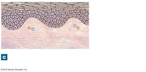
Cutaneous Membrane: Stratified squamous epithelium supported by dense irregular connective tissue; covers outer surface of the body.
Synovial Membranes: Areolar tissue, no epithelium; secrete and retain synovial fluid; found in articular capsules and bursae.

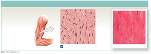
Muscle Tissue
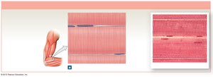
Skeletal Muscle Tissue
Cells (muscle fibers) are multinucleated and striated due to actin and myosin arrangement. Voluntary muscle controlled by CNS; responsible for skeletal movement and sphincters. Regenerates well.
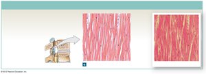
Cardiac Muscle Tissue
Cardiomyocytes are mono-nucleated, striated, and occur only in the heart. Involuntary muscle relying on pacemaker cells; interconnected by intercalated discs with gap junctions. Regenerates poorly.
*Additional info: Intercalated discs allow rapid propagation of action potentials for synchronized contraction.*
Smooth Muscle Tissue
Involuntary muscle lining hollow organs, driven by pacemaker cells and/or nerves. Non-striated due to different actin and myosin arrangement. Can divide and regenerate.
Nervous Tissue
Structure and Function
Nervous tissue contains neurons and neuroglia. Neurons convey information via electrical impulses, while neuroglia provide support.
*Additional info: Neurons consist of dendrites (receive signals), axons (transmit signals), and a cell body (contains nucleus). Neuroglia include astrocytes, oligodendrocytes, microglia, and ependymal cells.* ----------------------------------------
