 Back
BackChapter 4: Tissues – The Living Fabric (ANP Study Notes)
Study Guide - Smart Notes
 Tailored notes based on your materials, expanded with key definitions, examples, and context.
Introduction to Tissues
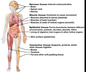
Tissues are groups of cells similar in structure that perform common or related functions. The study of tissues is called histology. There are four primary tissue types in the human body, each with distinct roles essential for maintaining homeostasis.
Epithelial tissue: Covers surfaces and forms boundaries.
Connective tissue: Supports, protects, and binds other tissues.
Muscle tissue: Produces movement.
Nervous tissue: Controls and communicates.
Epithelial Tissue
General Characteristics
Epithelial tissue forms boundaries between different environments, protects, secretes, absorbs, filters, and lines both external and internal surfaces. It is classified by location as either covering/lining epithelia or glandular epithelia.
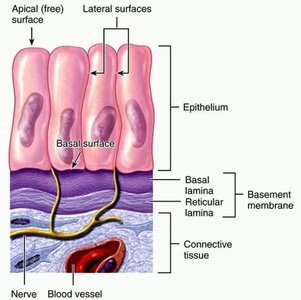
Polarity: Has an apical (free) surface and a basal (attached) surface, which differ in structure and function.
Specialized contacts: Cells fit closely together, forming continuous sheets bound by tight junctions and desmosomes.
Supported by connective tissue: The basement membrane (basal lamina + reticular lamina) reinforces the epithelium.
Avascular but innervated: Contains no blood vessels but is supplied by nerve fibers; nutrients diffuse from underlying tissues.
Regeneration: High regenerative capacity, especially when exposed to friction or hostile environments.
Classification of Epithelia
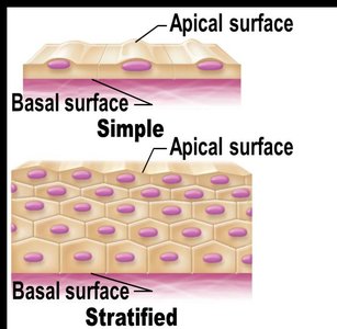
Epithelia are classified by the number of cell layers and the shape of the cells at the apical surface.
Simple epithelia: Single layer of cells.
Stratified epithelia: Two or more layers; named by the shape of cells in the apical layer.
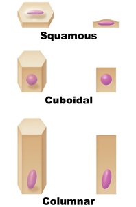
Cell shapes: Squamous (flat), cuboidal (boxlike), columnar (tall).
Types of Epithelial Tissue
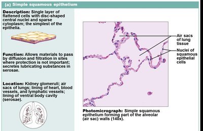
Simple Squamous Epithelium: Single layer of flat cells; allows diffusion and filtration. Locations: Air sacs of lungs, lining of heart, blood vessels, lymphatic vessels, serosae. Function: Absorption, diffusion, filtration, secretion.
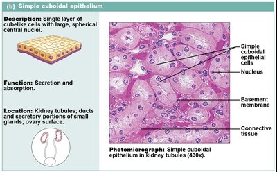
Simple Cuboidal Epithelium: Single layer of cube-shaped cells; secretion and absorption. Locations: Kidney tubules, ducts of small glands, ovary surface.
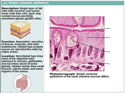
Simple Columnar Epithelium: Single layer of tall cells; absorption and secretion. Locations: Digestive tract, gallbladder, excretory ducts, small bronchi, uterine tubes.
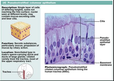
Pseudostratified Columnar Epithelium: Single layer of cells of differing heights; secretion, particularly of mucus. Locations: Trachea, upper respiratory tract, ducts of large glands.
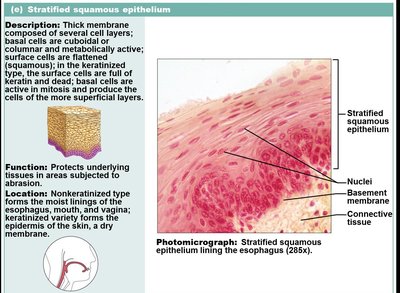
Stratified Squamous Epithelium: Several layers; protects underlying tissues. Locations: Nonkeratinized: mouth, esophagus, vagina; Keratinized: epidermis of skin.
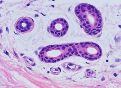
Stratified Cuboidal and Columnar Epithelium: Rare; secretion and protection. Locations: Sweat glands, mammary glands, pharynx, male urethra.
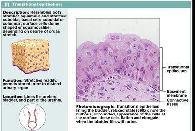
Transitional Epithelium: Resembles both stratified squamous and cuboidal; stretches readily. Locations: Lines ureters, bladder, part of urethra.
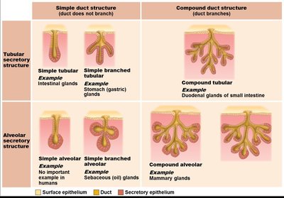
Glandular Epithelia
Glands are one or more cells that make and secrete an aqueous fluid called a secretion. They are classified by where they release their product (endocrine or exocrine) and the number of cells (unicellular or multicellular).
Endocrine glands: Ductless; secrete hormones into blood or lymph.
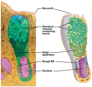
Exocrine glands: Secrete products into ducts onto body surfaces or cavities; can be unicellular (goblet cells) or multicellular.
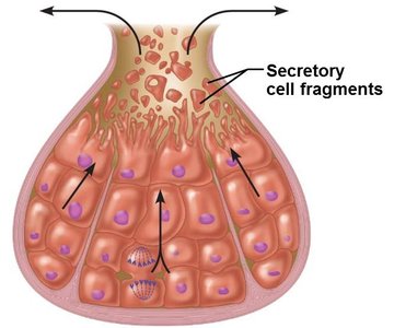
Merocrine: Secrete by exocytosis (e.g., sweat, salivary glands).
Holocrine: Accumulate products until cell ruptures (e.g., sebaceous glands).
Connective Tissue
General Characteristics and Functions
Connective tissue is the most abundant and widely distributed tissue type. It supports, protects, insulates, stores fuel, and transports substances. All connective tissues share three basic characteristics:
Derived from mesenchyme (embryonic tissue)
Varying degrees of vascularity
Composed largely of nonliving extracellular matrix (ground substance and fibers)
Structural Elements
Ground substance: Unstructured material that fills space between cells; contains interstitial fluid, cell adhesion proteins, and proteoglycans.
Fibers: Collagen (strength), elastic (stretch/recoil), reticular (supportive networks).
Cells: "Blast" cells (immature, matrix-secreting), "cyte" cells (mature, maintain matrix), fat cells, white blood cells, mast cells, macrophages.
Types of Connective Tissue
Connective Tissue Proper: Loose (areolar, adipose, reticular) and dense (regular, irregular, elastic).
Cartilage: Hyaline, elastic, fibrocartilage.
Bone (Osseous Tissue): Compact and spongy bone.
Blood: Fluid matrix (plasma), red and white blood cells.
Muscle Tissue
Muscle tissue is highly vascularized and responsible for movement. There are three types:
Skeletal muscle: Voluntary, attached to bones, striated, multinucleated.
Cardiac muscle: Involuntary, found in heart walls, striated, branched, intercalated discs.
Smooth muscle: Involuntary, found in walls of hollow organs, non-striated, spindle-shaped cells.
Nervous Tissue
Nervous tissue is specialized for internal communication. It consists of neurons (generate and conduct impulses) and neuroglia (supporting cells). Found in the brain, spinal cord, and nerves.
Covering and Lining Membranes
Cutaneous membrane: Skin; keratinized stratified squamous epithelium attached to dense irregular connective tissue (dermis); dry membrane.
Mucous membrane: Lines body cavities open to exterior; moist; epithelial sheet over lamina propria; may secrete mucus.
Serous membrane: Lines closed ventral body cavities; simple squamous epithelium (mesothelium) on areolar connective tissue; secretes serous fluid.
Tissue Repair
Tissue repair occurs in two major ways: regeneration (replacement with same tissue) and fibrosis (replacement with scar tissue). The process involves inflammation, organization (restoring blood supply), and regeneration/fibrosis.
Regenerative Capacity of Tissues
High: Epithelial tissues, bone, areolar connective tissue, dense irregular connective tissue, blood-forming tissue.
Moderate: Smooth muscle, dense regular connective tissue.
Weak: Skeletal muscle, cartilage.
None/Very limited: Cardiac muscle, nervous tissue in brain and spinal cord.
Developmental and Aging Aspects of Tissues
Primary germ layers: Ectoderm (nervous tissue), mesoderm (muscle, connective), endoderm (epithelial, plus others).
Aging: Epithelia thin, repair less efficient, tissues atrophy, increased cancer risk due to DNA mutations.
