 Back
BackChapter 4: Tissue—The Living Fabric (ANP Study Notes)
Study Guide - Smart Notes
 Tailored notes based on your materials, expanded with key definitions, examples, and context.
Tissues: Introduction and Overview
Definition and Classification of Tissues
Tissues are groups of cells with similar structure and function, organized to perform specific activities in the body. The arrangement and type of cells in a tissue determine its role, ranging from forming thin sheets to large masses. There are four primary tissue types in the human body:
Epithelial tissue: Forms sheets that cover or line body surfaces and cavities.
Connective tissue: Provides structural and functional support.
Muscle tissue: Contracts to produce movement.
Nervous tissue: Senses, conducts, and processes information.
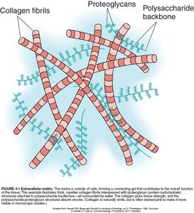
The matrix is the non-cellular component present within all tissues and organs, providing structural and biochemical support to the surrounding cells.
Epithelial Tissue
General Characteristics
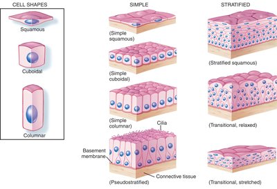
Epithelial tissue covers the body and lines body cavities. Its cells are tightly packed with minimal matrix, forming protective barriers and specialized surfaces for absorption, secretion, and filtration. Epithelial tissues are classified by both cell shape and arrangement.
Classification by Cell Shape
Squamous: Flat and scalelike
Cuboidal: Cube-shaped
Columnar: Taller than wide
Transitional: Varying shapes that can stretch
Classification by Cell Arrangement
Simple: Single layer of cells of the same shape
Stratified: Multiple layers of cells, named for the shape of cells in the outer layer
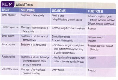
Types of Epithelial Tissues
Tissue | Structure | Locations | Functions |
|---|---|---|---|
Simple squamous | Single layer of flattened cells | Alveoli of lungs, lining of blood and lymphatic vessels | Diffusion of respiratory gases, filtration, osmosis |
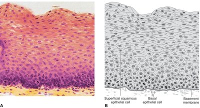
Stratified squamous | Many layers; outermost layer is flattened cells | Surface of lining of mouth and esophagus | Protection |
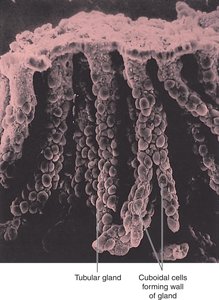
Simple cuboidal | Single layer of cube-shaped cells | Glands, kidney tubules, surface of skin (epidermis) | Secretion, absorption, protection |
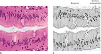
Simple columnar | Single layer of tall, narrow cells | Lining of stomach, intestines, parts of respiratory tract | Protection, secretion, transport (absorption) |
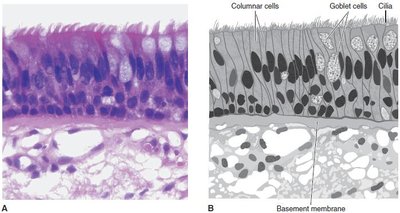
Pseudostratified | Single layer of tall cells that appear stratified | Lining of portions of the respiratory tract | Protection |
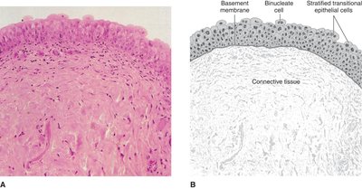
Stratified transitional | Many layers of varying shapes, capable of stretching | Urinary bladder | Protection |
Examples of Epithelial Tissues
Simple squamous epithelium: Single layer of flat cells; found in alveoli and blood vessels; allows for diffusion and filtration.
Stratified squamous epithelium: Multiple layers; found in skin, mouth, and esophagus; provides protection against abrasion.
Simple cuboidal epithelium: Single layer of cube-shaped cells; found in glands and kidney tubules; specialized for secretion and absorption.
Simple columnar epithelium: Single layer of tall cells; lines stomach and intestines; contains goblet cells for mucus production and is specialized for absorption.
Pseudostratified epithelium: Appears layered but all cells touch the basement membrane; lines the trachea; often ciliated for moving mucus.
Stratified transitional epithelium: Multiple layers of cells that can stretch; found in the urinary bladder.

Connective Tissue
General Characteristics
Connective tissue is the most abundant and widely distributed tissue type in the body. It consists of relatively few cells embedded in an extensive extracellular matrix, which determines the tissue's properties and functions. Connective tissue supports, binds, and protects other tissues and organs.
Types of Connective Tissue
Areolar: Loose arrangement of fibers; acts as a "glue" holding organs together.
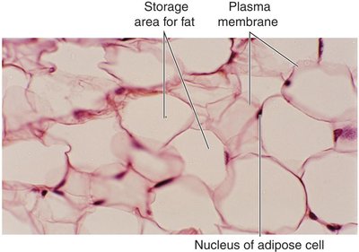
Adipose (fat): Stores lipids; insulates and protects organs.
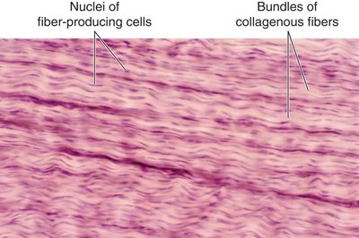
Fibrous: Dense bundles of collagen fibers; forms tendons and ligaments.
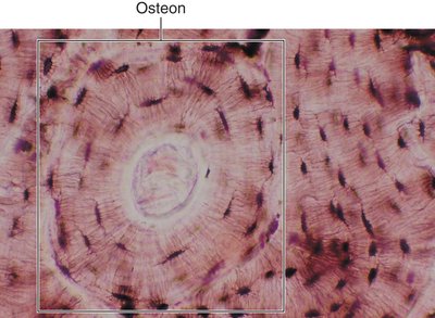
Bone: Calcified matrix; provides support and protection.
Cartilage: Gel-like matrix; provides flexible support; chondrocytes are the main cell type.
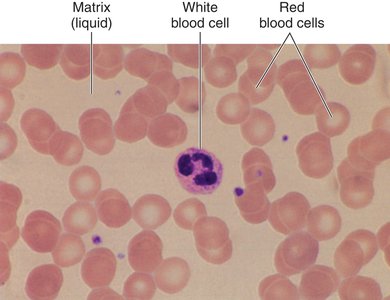
Blood: Fluid matrix (plasma); transports substances and provides immune protection.
Tissue | Structure | Locations | Functions |
|---|---|---|---|
Loose fibrous (areolar) | Loose arrangement of collagen fibers, elastic fibers, and cells | Area between other tissues and organs | Connection |
Adipose (white and brown fat) | Cells contain triglyceride vesicles | White fat: under skin, padding at various points; Brown fat: pockets within neck and torso | White fat: protection, insulation, support, nutrient reserve; Brown fat: heat production, regulation |
Dense fibrous (regular and irregular) | Dense arrangement of collagen fiber bundles forming parallel rows (regular) or irregular patterns | Tendons, ligaments, skin (deep layer), fascia, scar tissue | Flexible but strong connection |
Bone (compact and cancellous) | Hard, calcified matrix arranged in osteons (compact) or network of beams (cancellous) | Skeleton | Support, protection |
Cartilage (hyaline, fibrocartilage, elastic) | Hard but flexible matrix with embedded chondrocytes | Hyaline: nasal septum, larynx, trachea, bronchi, ends of bones; Fibrocartilage: disks between vertebrae, knee joint; Elastic: external ear | Hyaline: firm but flexible support; Fibrocartilage: withstands pressure; Elastic: flexible support |
Blood | Liquid matrix with flowing red and white cells | Blood vessels | Transportation, protection |
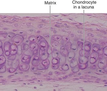
Major Types of Cartilage
Hyaline cartilage: Most common; found in nose, trachea, and at the ends of long bones.
Fibrocartilage: Strongest; found in intervertebral discs and knee menisci.
Elastic cartilage: Most flexible; found in the external ear.
Muscle Tissue
Introduction and Types
Muscle tissue is specialized for contraction, enabling movement, stability, and heat production. Muscle cells have a high degree of contractility and are classified into three types:
Skeletal muscle tissue: Attaches to bones; voluntary control; striated appearance.
Cardiac muscle tissue: Found in the heart wall; involuntary control; striated with intercalated disks.
Smooth muscle tissue: Found in walls of blood vessels and hollow organs; involuntary control; non-striated.
Nervous Tissue
Structure and Function
Nervous tissue is responsible for rapid communication and control of body functions. It consists of two main cell types:
Neurons: Conduct electrical impulses; each has a cell body, one axon (carries impulses away from the cell body), and one or more dendrites (carry impulses toward the cell body).
Glia (neuroglia): Supportive and connecting cells that assist neuron function.
Tissue Repair
Regeneration and Healing
Tissue repair is usually accomplished by regeneration, where new cells replace damaged ones. Epithelial and connective tissues regenerate easily, while muscle and nervous tissues have limited regenerative capacity. Scar tissue (fibrosis) may form if regeneration is incomplete.
Review Questions
Which tissue type can be subdivided according to the shape and arrangement of the cells found in each type? Answer: Epithelial
Name the type of epithelial tissue that is typically found in body areas subjected to stress and must be able to stretch. Answer: Stratified transitional
Tendons are composed of this type of connective tissue. Answer: Dense fibrous
Because its matrix is liquid, _____ is perhaps the most unusual form of connective tissue. Answer: Blood
The presence of intercalated disks is a unique characteristic of which type of muscle tissue? Answer: Cardiac
Which type of tissue functions in rapid communication between body structures and controls body functions? Answer: Nervous
Which tissue type has a limited capacity to regenerate? Answer: Muscle
