 Back
BackChapter 4: Tissue—The Living Fabric (Epithelial and Connective Tissues)
Study Guide - Smart Notes
 Tailored notes based on your materials, expanded with key definitions, examples, and context.
Tissue: The Living Fabric
Introduction to Tissues
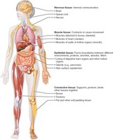
Tissues are groups of cells similar in structure that perform common or related functions, maintaining homeostasis in the body. The study of tissues is called histology. There are four basic tissue types: epithelial, connective, muscle, and nervous tissue.
Epithelial Tissue
Definition and Functions
Epithelial tissue (epithelium) is a sheet of cells that covers body surfaces or lines body cavities. It has two main forms: covering and lining epithelium (e.g., skin, lining of digestive tract) and glandular epithelium (e.g., salivary glands). Main functions include protection, absorption, filtration, excretion, secretion, and sensory reception.
Special Characteristics of Epithelial Tissues
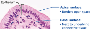
Polarity: Cells have an apical (top, exposed) and basal (bottom, attached) surface. The apical surface may have microvilli for absorption. The basal surface attaches to the basal lamina, anchoring the epithelium to underlying tissues.
Specialized Contacts: Epithelial cells fit closely together, forming continuous sheets. Lateral contacts include tight junctions and desmosomes.
Supported by Connective Tissue: All epithelial sheets rest upon and are supported by connective tissue (basement membrane).
Avascular but Innervated: Epithelia lack blood vessels but are supplied by nerve fibers; nutrients diffuse from underlying tissues.
Regeneration: Epithelial cells have high regenerative capacity, especially in areas exposed to friction or hostile environments.
Classification of Epithelial Tissue
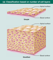
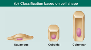
Epithelia are classified by the number of cell layers and cell shape:
Simple epithelia: Single cell layer (for absorption, secretion, filtration).
Stratified epithelia: Two or more layers (for protection in high-abrasion areas).
Cell shapes: Squamous (flattened), cuboidal (cube-like), columnar (tall).
Types of Epithelial Tissue
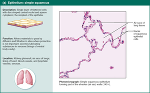
Simple Squamous Epithelium
Single layer of flattened cells; allows rapid diffusion and filtration. Found in kidney glomeruli, air sacs of lungs, lining of heart, blood vessels, and lymphatic vessels. Special types include endothelium (lining vessels) and mesothelium (serous membranes).
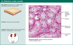
Simple Cuboidal Epithelium
Single layer of cube-like cells; functions in secretion and absorption. Located in kidney tubules, ducts, and secretory portions of small glands.
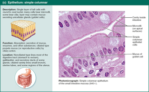
Simple Columnar Epithelium
Single layer of tall, closely packed cells; may have microvilli or cilia. Functions in absorption and secretion of mucus, enzymes, and other substances. Found in digestive tract, gallbladder, ducts of some glands, bronchi, and uterine tubes.
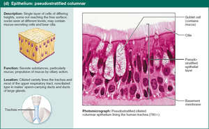
Pseudostratified Columnar Epithelium
Single layer of cells of varying heights, giving a false impression of stratification. Often ciliated; functions in secretion and movement of mucus. Located in upper respiratory tract, ducts of large glands, and tubules in testes.
Stratified Epithelia (Overview)
Stratified epithelia involve two or more layers of cells, providing protection in high-wear areas (e.g., skin, mouth lining). The most widespread type is stratified squamous epithelium. Other types include stratified cuboidal and columnar (rare), and transitional epithelium (lines urinary organs).
*Additional info: For further details on stratified and transitional epithelia, refer to textbook figures and histology atlases.*
