 Back
BackChapter 4A: Tissue – The Living Fabric (Epithelial Tissue Study Guide)
Study Guide - Smart Notes
 Tailored notes based on your materials, expanded with key definitions, examples, and context.
Tissue: The Living Fabric
Introduction to Tissues
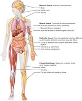
Tissues are groups of cells with similar structure and function, essential for maintaining homeostasis in the human body. The study of tissues is known as histology. There are four basic tissue types: epithelial, connective, muscle, and nervous tissue.
Epithelial tissue: Covers surfaces, lines cavities, forms glands.
Connective tissue: Supports, protects, binds other tissues.
Muscle tissue: Contracts to cause movement.
Nervous tissue: Enables internal communication.
Histological Preparation of Tissue Samples
To study tissues under a microscope, samples must be properly prepared. This process involves:
Fixation: Preserving tissue with a solvent to prevent decay.
Sectioning: Cutting tissue into thin slices for light or electron transmission.
Staining: Applying dyes or heavy metal salts to enhance contrast. Light microscopy uses colored dyes; electron microscopy uses heavy metal salts.
Artifacts may occur, causing distortions from the living state. Transmission electron microscopy (TEM) shows internal sections, while scanning electron microscopy (SEM) reveals surface details.
Epithelial Tissue
General Characteristics and Functions
Epithelial tissue (epithelium) is a sheet of cells covering body surfaces or lining cavities. It exists in two main forms:
Covering and lining epithelium: Forms the skin's outer layer, lines open cavities, and covers organs.
Glandular epithelium: Forms glands such as salivary glands.
Main functions include protection, absorption, filtration, excretion, secretion, and sensory reception.
Special Characteristics of Epithelial Tissue
Epithelial tissues possess five key characteristics:
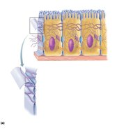
Polarity: Cells have distinct apical (top) and basal (bottom) surfaces. The apical surface is exposed to the cavity or external environment, often featuring microvilli. The basal surface attaches to the basal lamina, anchoring the epithelium to underlying tissues.

Specialized contacts: Cells are tightly joined by lateral contacts such as tight junctions and desmosomes, forming continuous sheets.

Supported by connective tissue: All epithelial sheets rest on connective tissue, with the basement membrane (basal and reticular lamina) providing structural support and boundary definition.

Avascular but innervated: Epithelial tissues lack blood vessels and receive nutrients by diffusion, but are supplied with nerve fibers.
Regeneration: Epithelial cells have high regenerative capacity, replacing damaged cells rapidly if nutrients are available.
Classification of Epithelial Tissue
Epithelial tissues are classified by two criteria:
Number of cell layers:
Simple epithelium: Single cell layer, ideal for absorption, secretion, filtration.
Stratified epithelium: Multiple layers, suited for protection in high-abrasion areas.

Cell shape:
Squamous: Flattened, scale-like.
Cuboidal: Box-like, cube-shaped.
Columnar: Tall, column-like.
In stratified epithelia, cell shape is named according to the apical layer.

Types of Epithelial Tissue
Simple Squamous Epithelium
Single layer of flattened cells with sparse cytoplasm. Functions in rapid diffusion and filtration. Found in kidney glomeruli, air sacs of lungs, lining of heart, blood vessels, and lymphatic vessels.
Endothelium: Lines lymphatic vessels, blood vessels, and heart.
Mesothelium: Forms serous membranes in ventral body cavity.

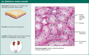
Simple Cuboidal Epithelium
Single layer of cube-shaped cells. Functions in secretion and absorption. Forms walls of small ducts of glands and kidney tubules.
Simple Columnar Epithelium
Single layer of tall, closely packed cells. Some have microvilli or cilia; goblet cells secrete mucus. Functions in absorption and secretion of mucus, enzymes, and other substances. Ciliated cells move mucus. Found in digestive tract, gallbladder, ducts of glands, bronchi, and uterine tubes.
Pseudostratified Columnar Epithelium
Appears multi-layered but is actually a single layer. Many cells are ciliated. Functions in secretion and movement of mucus. Located in upper respiratory tract, ducts of large glands, and tubules in testes.
Stratified Squamous Epithelium
Multiple layers; basal cells divide and migrate toward surface. Provides protection in high wear and tear areas. Most widespread stratified epithelium. Keratinized cells are found in skin; nonkeratinized in moist linings.
Stratified Cuboidal and Columnar Epithelium
Rare types. Stratified cuboidal found in sweat and mammary glands (usually two layers). Stratified columnar found in pharynx, male urethra, and some glandular ducts; usually at transition areas.
Transitional Epithelium
Lines hollow urinary organs (bladder, ureters, urethra). Basal cells are cuboidal or columnar. Cells change shape when stretched, allowing for increased urine flow and storage.
Glandular Epithelia
A gland is one or more cells that make and secrete an aqueous fluid called a secretion. Glands are classified by:
Site of product release:
Endocrine: Ductless, release hormones into interstitial fluid and blood.
Exocrine: Secrete products onto body surfaces or into cavities via ducts (e.g., sweat, oil, salivary glands).
Number of cells:
Unicellular: Goblet cells, mucous cells.
Multicellular: Salivary glands.
Formation of Multicellular Exocrine and Endocrine Glands
Multicellular glands develop from epithelial tissue, forming either ducts (exocrine) or losing ducts (endocrine).
Unicellular Exocrine Glands
Goblet cells and mucous cells produce mucin, which dissolves in water to form mucus—a protective, lubricating coating.
Multicellular Exocrine Glands
Composed of a duct and secretory unit, surrounded by connective tissue. Classified by structure and mode of secretion:
Structure:
Simple: Unbranched ducts.
Compound: Branched ducts.
Tubular: Secretory cells form tubes.
Alveolar: Secretory cells form sacs.
Tubuloalveolar: Both types present.
Modes of Secretion
Merocrine: Secrete by exocytosis as produced; most common (sweat, pancreas, salivary glands).
Holocrine: Accumulate products until cell ruptures; replaced by division (sebaceous glands).
Apocrine: Apex of cell ruptures; cell repairs and repeats. Controversial in humans; mammary glands are closest example.
Summary Table: Epithelial Tissue Classification
Type | Structure | Function | Location |
|---|---|---|---|
Simple Squamous | Single layer, flat | Diffusion, filtration | Kidney, lungs, vessels |
Simple Cuboidal | Single layer, cube | Secretion, absorption | Kidney tubules, glands |
Simple Columnar | Single layer, tall | Absorption, secretion | Digestive tract, ducts |
Pseudostratified Columnar | Single layer, varied height | Mucus secretion, movement | Respiratory tract |
Stratified Squamous | Multiple layers, flat apical | Protection | Skin, mouth, esophagus |
Stratified Cuboidal | 2+ layers, cube | Protection | Sweat, mammary glands |
Stratified Columnar | 2+ layers, column apical | Protection, secretion | Pharynx, urethra |
Transitional | Multiple layers, shape varies | Stretching | Bladder, ureters |
Additional info: This guide expands on brief lecture points with academic context, definitions, and examples for clarity and completeness.
