 Back
Chapter 5: The Integumentary System – High-Yield Study Guide
Study Guide - Smart Notes
 Tailored notes based on your materials, expanded with key definitions, examples, and context.
The Integumentary System
Overview
The integumentary system, primarily composed of the skin, is the largest organ system in the body. It serves as a protective barrier, regulates temperature, and houses sensory receptors. The skin consists of three main layers: the epidermis, dermis, and hypodermis (subcutaneous tissue).
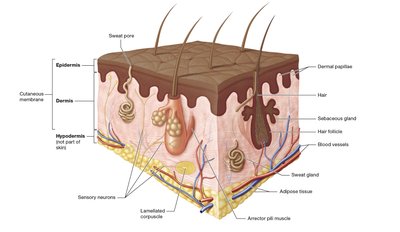
Epidermal Structure & Cells
The Five Layers of the Epidermis
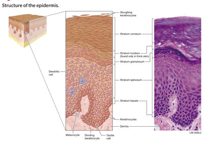
The epidermis is avascular and relies on diffusion from the underlying dermis. It is composed of stratified squamous epithelial tissue and organized into five distinct layers (from deep to superficial):
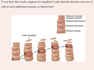
Stratum Basale: Single row of actively dividing stem cells; closest to the blood supply.
Stratum Spinosum: Thickest living layer; cells appear spiny due to desmosomes.
Stratum Granulosum: Cells release hydrophobic lipids, forming a water barrier and leading to apoptosis.
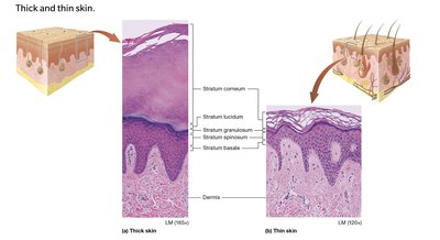
Stratum Lucidum: Clear, dead layer found only in thick skin (palms, soles).
Stratum Corneum: Multiple layers of dead, flattened, keratinized cells that are shed daily.
Core Cell Types in the Epidermis
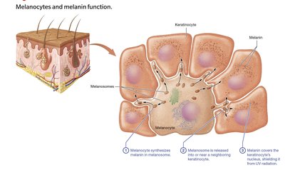
Keratinocytes: Make up 95% of epidermal cells; produce keratin for strength and are connected by desmosomes.
Melanocytes: Located in the stratum basale; produce melanin in melanosomes, which protect DNA from UV radiation.
Tactile (Merkel) Cells: Also in the stratum basale; function as light touch receptors.
Dendritic (Langerhans) Cells: Found in the stratum spinosum; act as immune phagocytes.
Dermal Layers & Skin Markings
Dermal Structure
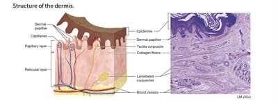
The dermis is highly vascular and composed of two layers:
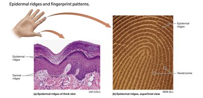
Papillary Layer: Areolar connective tissue; contains dermal papillae (wavy folds with capillary loops) and Meissner corpuscles (light touch receptors). Forms friction ridges (fingerprints).
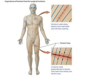
Reticular Layer: Dense irregular connective tissue; contains collagen and elastic fibers, Pacinian corpuscles (deep pressure/vibration receptors), flexure lines, and tension lines (cleavage lines).
Clinical Note: Blisters and Stretch Marks
Blisters: Fluid-filled separation between epidermis and dermis due to friction or burns.
Striae (Stretch Marks): Tearing of dermal elastic fibers from rapid stretching.
Skin Pigmentation & Clinical Clues
Determinants of Skin Color
Skin color is determined by the interaction of three main pigments:
Melanin: Orange-red to black pigment produced by melanocytes; everyone has the same number of melanocytes, but production varies.
Carotene: Yellow-orange pigment from diet; accumulates in the stratum corneum.
Hemoglobin: Bright red blood protein; gives a pink hue to fair skin.
Pathological Discoloration Cheat Sheet
Cyanosis: Bluish tint from oxygen-starved hemoglobin; indicates respiratory distress or shock.
Erythema: Redness from increased blood flow due to exercise, fever, or inflammation.
Pallor: Pale appearance from decreased blood flow, revealing white dermal collagen.
Jaundice: Yellowish tint from bilirubin accumulation, indicating liver dysfunction.
Accessory Structures: Hair, Nails, and Glands
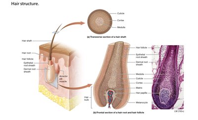
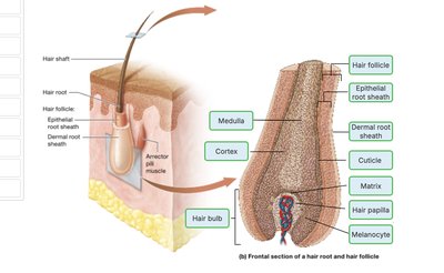
Hair Structure and Function
Hair Layers: Medulla (core) → Cortex (middle) → Cuticle (outer layer).
Hair Matrix: Growth zone at the base of the follicle; site of rapid mitosis.
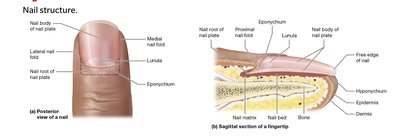
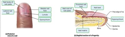
Nail Structure and Function
Nail Matrix: Hidden growth zone under the root; produces the nail plate.
Lunula: Opaque, white crescent at the nail base.
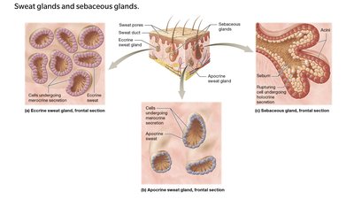
Cutaneous Glands
Type | Secretion | Location | Function |
|---|---|---|---|
Eccrine Sweat | Watery sweat | Most skin | Thermoregulation, antimicrobial protection |
Apocrine Sweat | Protein-rich sweat | Axillae, groin | Body odor (bacterial action), activated at puberty |
Sebaceous | Sebum (oil) | Face, scalp (not palms/soles) | Moisture seal, antibacterial, lubricates hair |
Pathology: Burns & Skin Cancer
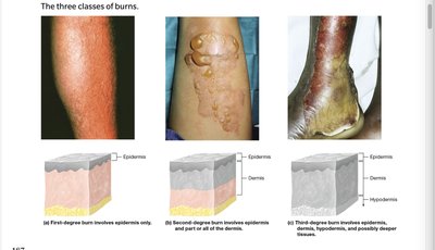
Burn Depth Classification
1st-Degree: Epidermis only; redness and pain, no blisters.
2nd-Degree: Epidermis + part of dermis; blisters and intense pain.
3rd-Degree: Epidermis, dermis, and hypodermis; painless (nerve destruction), severe scarring.
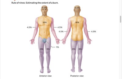
Rule of Nines
Used to estimate the percentage of body surface area burned for fluid resuscitation:
Skin Cancer Profiles
Basal Cell Carcinoma: Most common; arises from stratum basale keratinocytes; rarely metastasizes.
Squamous Cell Carcinoma: From stratum spinosum keratinocytes; may ulcerate and bleed.
Malignant Melanoma: Most dangerous; arises from melanocytes; highly metastatic.
ABCDE Framework for Melanoma Detection
Asymmetry: Two halves do not match.
Border: Irregular, ragged, or blurred edges.
Color: Uneven shades or unusual colors.
Diameter: Greater than 6 mm.
Evolving: Changes in size, shape, or color.
Additional info:
The hypodermis (subcutaneous layer) is not technically part of the skin but anchors it to underlying tissues and stores fat.
Incisions made parallel to tension lines heal with less scarring due to the orientation of collagen fibers.
