 Back
BackChapter 5: The Integumentary System – Structure, Function, and Clinical Relevance
Study Guide - Smart Notes
 Tailored notes based on your materials, expanded with key definitions, examples, and context.
The Integumentary System
Overview
The integumentary system is composed of the skin and its accessory structures, including hair, nails, and glands. It serves as the body's primary barrier against environmental hazards and plays a crucial role in homeostasis, protection, and sensory perception. 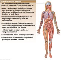
Protection: Shields internal tissues from mechanical damage, pathogens, and dehydration.
Temperature Regulation: Maintains normal body temperature via sweat and blood flow.
Sensory Reception: Detects touch, pressure, pain, and temperature.
Vitamin D3 Synthesis: Initiates production of vitamin D3 in the epidermis under UV exposure.
Excretion: Removes salts, water, and organic wastes.
Vitamin D3 and the Skin
Production and Function
Vitamin D3 (cholecalciferol) is synthesized by epidermal cells when exposed to ultraviolet (UV) radiation. It is essential for calcium and phosphate absorption in the small intestine, supporting bone health. 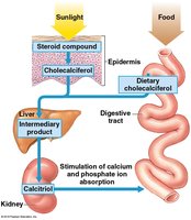
Synthesis: UV light converts a steroid compound in the epidermis to cholecalciferol.
Conversion: The liver and kidneys convert cholecalciferol to calcitriol, the active form.
Function: Calcitriol stimulates absorption of calcium and phosphate ions.
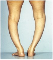
Deficiency: Insufficient vitamin D3 can cause rickets, a disease characterized by weak, flexible bones.
Clinical Example: Rickets
Rickets: Results from vitamin D3 deficiency, leading to bone deformities and structural changes.
Hair and Hair Follicles
Structure and Function
Hair and its associated structures are derived from the embryonic epidermis and are located in the dermis, projecting to the skin surface.
Functions: Protection, insulation, guarding openings, and sensory reception.
Distribution: Hairs cover most of the body except palms, soles, lips, and parts of external genitalia.
Hair Follicle Anatomy
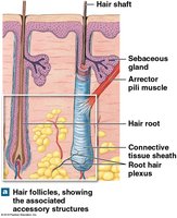
Hair Follicle: Extends deep into the dermis, produces nonliving hairs, and is wrapped in a connective tissue sheath.
Arrector Pili Muscle: Involuntary smooth muscle causing "goose bumps" by raising hairs.
Root Hair Plexus: Sensory nerves surrounding the base of the follicle.
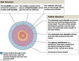
Hair Structure
Medulla: Central core containing soft keratin.
Cortex: Intermediate layer with hard keratin for stiffness.
Cuticle: Surface layer with hard keratin.
Follicle Layers
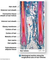
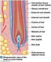
Internal Root Sheath: Surrounds hair root.
External Root Sheath: Extends from skin surface to hair matrix.
Glassy Membrane: Clear layer wrapped in connective tissue sheath.
Hair Growth and Types
Hair Production: Begins at the hair bulb, surrounding the hair papilla (capillaries and nerves).
Hair Matrix: Epithelial cells divide, pushing hair up and out.
Growth Cycle: Includes active growth, club hair formation, and shedding.
Types: Vellus hairs (soft, fine) and terminal hairs (heavy, pigmented).
Hair Color: Determined by melanocytes at the hair matrix and genetic factors.
Sebaceous and Sweat Glands
Sebaceous (Oil) Glands
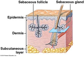
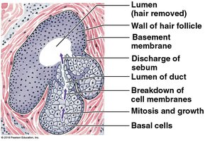
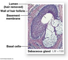
Holocrine Glands: Discharge sebum into hair follicles.
Functions: Lubricates and protects hair shaft, inhibits bacterial growth.
Types: Simple branched alveolar glands (with hair) and sebaceous follicles (without hair).
Sweat Glands
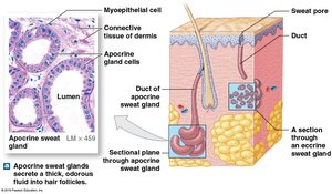
Apocrine Sweat Glands: Found in armpits, nipples, and pubic region; secrete into hair follicles via merocrine secretion; produce sticky, cloudy secretions; nutrient source for bacteria (odor).
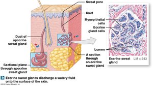
Eccrine (Merocrine) Sweat Glands: Widely distributed; discharge directly onto skin surface; secretions are 99% water; functions include cooling, excretion, and protection.
Other Integumentary Glands
Mammary Glands: Produce milk.
Ceruminous Glands: Produce cerumen (earwax), protecting the eardrum.
Control of Glands
Autonomic Nervous System (ANS): Controls sebaceous and apocrine glands globally.
Eccrine Glands: Controlled locally for thermoregulation.
Thermoregulation: Eccrine glands and cardiovascular system regulate body temperature.
Nails
Anatomical Structure and Formation
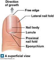
Nails protect the tips of fingers and toes and are composed of dead cells packed with keratin.
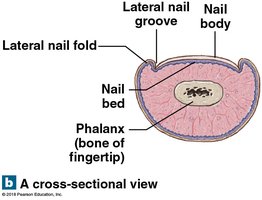
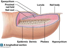
Nail Body: Visible portion covering the nail bed.
Lateral Nail Grooves and Folds: Surround the sides of the nail.
Hyponychium: Skin beneath the distal free edge.
Nail Root: Site of nail production, close to bone.
Eponychium: Cuticle, visible nail emerges from under nail root.
Lunula: Pale crescent near the root due to obscured blood vessels.
Repair of the Integument
Response to Injury
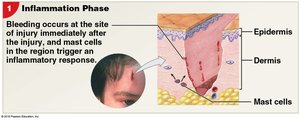
The skin responds to injury through a series of steps involving inflammation, tissue repair, and scar formation.
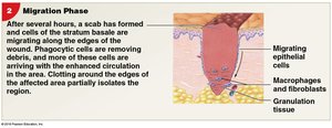
Inflammation: Bleeding, swelling, pain; mast cells trigger response.
Scab Formation: Stabilizes and protects the area.
Macrophages: Clean the area.
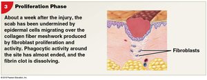
Granulation Tissue: Produced by fibroblasts and endothelial cells.
Scar Formation
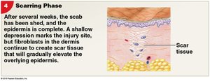
Clot Dissolution: Capillaries decline, noncellular scar tissue forms.
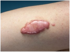
Keloid: Raised fibrous scar tissue may develop.
Aging and the Integumentary System
Effects of Aging
Aging leads to structural and functional changes in the skin, affecting its protective and regulatory roles.
Epidermis Thins: Increased vulnerability to injury.
Dendritic Cells Decrease: Reduced immune response.
Vitamin D3 Production Declines: Impacts bone health.
Melanocyte and Glandular Activity Declines: Less pigmentation and moisture.
Blood Supply to Dermis Reduces: Slower healing.
Hair Follicle Function Declines: Hair loss.
Elastic Fiber Network Shrinks: Reduced skin elasticity.
Sex-Specific Hair and Fat Distribution Fades: Changes in appearance.
Repair Rate Slows: Longer recovery from injury.
