 Back
Chapter 5: The Integumentary System – Structured Study Notes
Study Guide - Smart Notes
 Tailored notes based on your materials, expanded with key definitions, examples, and context.
The Integumentary System
Overview
The integumentary system is the body's largest organ system, consisting of the skin and its appendages (hair, nails, sweat glands, and sebaceous glands). It serves as a protective barrier and plays vital roles in sensation, thermoregulation, and metabolic functions.
Components: Skin, hair, nails, sweat glands, sebaceous (oil) glands
Main Functions: Protection, regulation, sensation, metabolic activity, blood reservoir, excretion
Skin Structure
Layers of the Skin
The skin is composed of three primary layers: the epidermis, dermis, and hypodermis (subcutaneous tissue). Each layer has distinct structural and functional characteristics.
Epidermis: Outermost layer; keratinized stratified squamous epithelium
Dermis: Middle layer; strong, flexible connective tissue
Hypodermis: Deepest layer; adipose tissue for insulation and energy storage
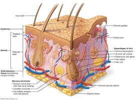
Epidermis
The epidermis is a keratinized stratified squamous epithelium containing four main cell types and organized into distinct layers (strata).
Keratinocytes: Most abundant; produce keratin for protection
Melanocytes: Produce melanin pigment; protect DNA from UV damage
Dendritic (Langerhans) Cells: Immune defense; detect and fight pathogens
Tactile (Merkel) Cells: Sensory receptors; detect touch
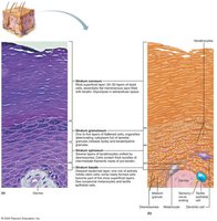
Layers of the Epidermis
The epidermis consists of 4-5 layers, depending on skin thickness:
Stratum basale: Deepest; single row of stem cells; site of cell division
Stratum spinosum: Several layers; strong connections; contains prekeratin fibers
Stratum granulosum: Keratinization begins; cells start to die; water-resistant barrier
Stratum lucidum: Only in thick skin; clear, flat, dead keratinocytes
Stratum corneum: Outermost; 20-30 layers of dead, keratinized cells; protection
Dermis
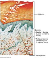
The dermis is a strong, flexible connective tissue layer containing fibroblasts, macrophages, blood vessels, nerves, and skin appendages. It is divided into two layers:
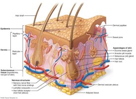
Papillary layer: Loose areolar connective tissue; contains dermal papillae, capillaries, nerve endings, and touch receptors
Reticular layer: Dense irregular connective tissue; provides strength and elasticity; contains collagen and elastic fibers, blood vessels, and some adipose tissue
Dermal Modifications
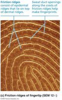
Dermal papillae: Fingerlike projections; form friction ridges in thick skin (fingerprints)
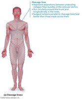
Cleavage (tension) lines: Parallel collagen fibers; important for surgical incisions
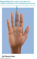
Flexure lines: Dermal folds near joints; visible creases in hands, wrists, fingers, soles, toes
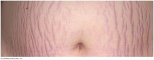
Striae (stretch marks): Silvery scars from dermal tearing
Blisters: Fluid-filled pockets separating epidermis and dermis
Skin Color
Pigments Contributing to Skin Color
Skin color is determined by three main pigments:
Melanin: Produced by melanocytes; shields DNA from UV; two forms (reddish yellow to brownish black)
Carotene: Yellow-orange pigment; stored in stratum corneum and hypodermis
Hemoglobin: Red pigment in blood; gives pinkish hue in lighter skin
Effects of UV Exposure
UV damages skin structure, increases cancer risk, alters DNA, suppresses immune system, destroys folic acid
Photosensitivity: Increased reaction to sunlight due to drugs or perfumes
Disease States and Skin Color
Cyanosis: Blue skin; low oxygenation
Pallor: Pale color; anemia, low blood pressure, fear, anger
Erythema: Redness; fever, hypertension, inflammation, allergy
Jaundice: Yellow cast; liver disorders
Bruises: Clotted blood under skin; color changes as blood breaks down
Dark "necklace" discoloration: Hyperpigmented areas; may indicate insulin resistance
Hair
Structure and Function
Hair is made of dead, keratinized cells and serves protective and sensory functions. It is not found on palms, soles, lips, nipples, or parts of external genitalia.
Functions: Detects insects, protects from trauma, reduces heat loss, shields from sunlight
Regions: Shaft (above skin), root (below skin)
Hair Shaft Structure
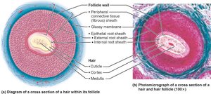
Medulla: Central core of large cells and air spaces
Cortex: Several layers of flattened cells surrounding medulla
Cuticle: Outer layer of overlapping single cells
Hair Color
Determined by melanocytes in hair follicles
Pheomelanin gives red tones
Gray/white hair: Decreased melanin, air bubbles replace pigment
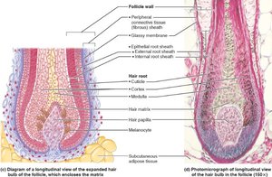
Structure of a Hair Follicle
Hair follicle: Extends from epidermis to dermis
Hair bulb: Expanded base; site of hair growth
Root hair plexus: Sensory nerve endings
Follicle wall layers: Fibrous sheath, glassy membrane, epithelial root sheath
Hair matrix: Actively dividing cells
Hair papilla: Capillaries supply nutrients
Arrector pili muscle: Smooth muscle; causes goosebumps
Types and Growth of Hair
Vellus hair: Fine, pale body hair
Terminal hair: Coarse, thick hair (scalp, eyebrows, axillary, pubic regions)
Growth regulation: Hormones and nutrition
Growth cycle: Growth and rest phases; grows ~2.25 mm/week; lose ~90 hairs/day
Hair Disorders
Hirsutism: Excess hair growth due to increased androgens
Alopecia: Hair thinning; can be genetic or hormone-influenced
Telogen effluvium: Sudden hair thinning; often due to stress, medications, hypothyroidism, low-protein diets
Nails
Structure and Function
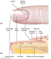
Nails are modified epidermis made of hard keratin, protecting the tips of fingers and toes.
Structure: Free edge, nail plate, root, nail bed, nail matrix (growth region)
Nail folds: Skin overlapping nail edges
Eponychium (cuticle): Protective seal at base
Hyponychium: Area under free edge
Lunule: White crescent; part of nail matrix
Nails appear pink due to capillaries
Nail Indicators
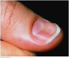
Yellow nails: Respiratory or thyroid disorders; thickening may indicate fungal infection
Koilonychia (spoon nails): Curved outward; may indicate iron deficiency
Beau’s lines: Horizontal grooves; linked to severe illness or stress
Cutaneous Glands
Sweat (Sudoriferous) Glands
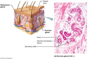
Sweat glands are present on most skin surfaces and are classified as eccrine (merocrine) or apocrine.
Eccrine (merocrine): Most numerous; found on palms, soles, forehead; open directly to skin pores; function in thermoregulation
Apocrine: Located in axillary and anogenital regions; secrete thick, milky sweat; ducts open into hair follicles; become active at puberty; may function as scent glands
Ceruminous glands: Ear canal; secrete cerumen (earwax)
Mammary glands: Produce milk
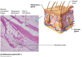
Sebaceous (Oil) Glands
Found throughout the body except palms and soles
Usually empty into hair follicles
Become active at puberty; stimulated by androgens
Secrete sebum (oily substance); lubricates skin and hair; antibacterial properties
Holocrine secretion: Cells rupture to release contents
Summary Table: Cutaneous Glands
Gland Type | Location | Secretion | Function |
|---|---|---|---|
Eccrine | Palms, soles, forehead | Watery sweat | Thermoregulation |
Apocrine | Axillary, anogenital | Milky sweat | Scent, body odor |
Ceruminous | Ear canal | Cerumen (earwax) | Protects ear |
Mammary | Breasts | Milk | Nourishment |
Sebaceous | All except palms/soles | Sebum (oil) | Lubrication, antibacterial |
Functions of Skin
Protection
The skin acts as a barrier against microorganisms, abrasions, temperature extremes, and chemicals. It provides three types of barriers:
Chemical barrier: Sweat, sebum, acid mantle, melanin
Physical barrier: Stratum corneum, lipids; blocks water and most substances
Biological barrier: Dendritic cells, macrophages, DNA protection
Body Temperature Regulation
Insensible perspiration: ~500 mL/day; not noticeable
Sensible perspiration: Up to 12 L/day; occurs with increased body temperature
Heat response: Vasodilation increases heat loss
Cold response: Vasoconstriction reduces heat loss
Cutaneous Sensations
Exteroceptors: Detect external stimuli (touch, temperature)
Free nerve endings: Detect pain
Metabolic Functions
Vitamin D synthesis: Needed for calcium absorption
Chemical protection: Neutralizes some carcinogens
Hormone activation: Converts cortisone to hydrocortisone
Collagen maintenance: Produces collagenase
Blood Reservoir
Skin can hold up to 5% of the body’s total blood volume
Vessels can be constricted to shunt blood to other organs
Excretion
Skin can secrete limited amounts of nitrogenous wastes (ammonia, urea, uric acid)
Sweating causes salt and water loss
Skin Cancer and Burns
Skin Cancer
Skin cancer is a major concern due to UV exposure and chronic irritation. There are three main types:
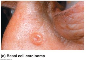
Basal cell carcinoma: Least malignant, most common; arises from stratum basale; cured by excision
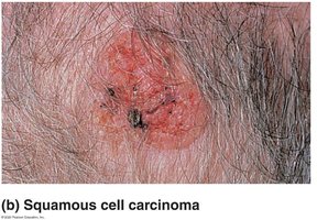
Squamous cell carcinoma: Second most common; can metastasize; arises from stratum spinosum
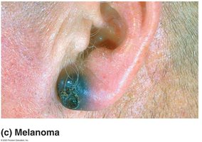
Melanoma: Cancer of melanocytes; most dangerous; highly metastatic; resistant to chemotherapy
Burns
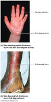
Burns are caused by heat, electricity, radiation, or chemicals. They result in protein denaturation and cell death, with dehydration and electrolyte imbalance as major dangers.
First-degree: Epidermis only; redness, swelling, pain
Second-degree: Epidermis + upper dermis; blisters present
Third-degree: Entire skin thickness; gray-white, red, or blackened; no pain (nerve endings destroyed); often requires skin grafting
Rule of Nines
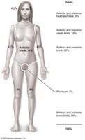
The Rule of Nines is used to estimate the extent and severity of burns by dividing the body into percentage regions.
Burns: Critical Criteria and Treatment
Burns are considered critical if: >25% of body has second-degree burns, >10% has third-degree burns, or face/hands/feet bear third-degree burns
Treatment: Debridement, antibiotics, temporary covering, skin grafts
