 Back
BackChapter 5: The Integumentary System – Structured Study Notes
Study Guide - Smart Notes
 Tailored notes based on your materials, expanded with key definitions, examples, and context.
Fundamentals of Anatomy & Physiology
Chapter 5: The Integumentary System
The integumentary system is the largest organ system in the human body, serving as the primary barrier between internal structures and the external environment. It consists of the cutaneous membrane (skin) and various accessory structures, each with specialized functions.
Introduction to the Integumentary System
Components of the Integumentary System
Cutaneous Membrane: Composed of the epidermis (superficial epithelium) and dermis (deep connective tissue).
Accessory Structures: Includes hair, hair follicles, exocrine glands, and nails.
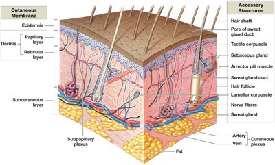
Subcutaneous Layer (Hypodermis)
Location: Deep to the dermis; separates skin from underlying organs.
Composition: Loose connective tissue, primarily adipose tissue.
Function: Stabilizes skin position, stores energy, and provides insulation.
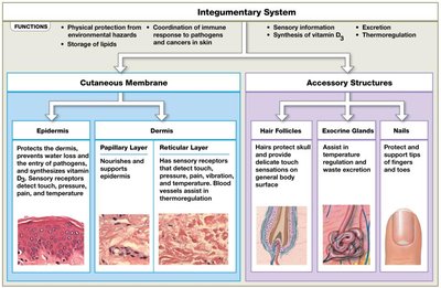
Functions of the Integumentary System
Protection: Shields underlying tissues from impact, abrasion, fluid loss, and microorganisms.
Excretion: Removes salts, water, and organic waste via exocrine glands.
Temperature Regulation: Maintains body temperature through insulation and evaporative cooling.
Melanin Production: Protects against UV radiation.
Keratin Production: Provides toughness and water resistance.
Vitamin D3 Synthesis: Essential for calcium metabolism.
Structure of the Skin
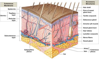
Layers of the Cutaneous Membrane
Epidermis: Keratinized, stratified squamous epithelium; avascular.
Dermis: Connective tissue with blood vessels and nerves; anchors accessory structures.
Hypodermis: Not part of the skin, but stabilizes its position.
Epidermis
Cell Types and Structure
Keratinocytes: Main cell type; produce keratin, a tough, fibrous protein.
Melanocytes: Produce melanin pigment.
Tactile (Merkel) Cells: Sensory receptors for touch.
Dendritic (Langerhans) Cells: Immune defense.
Types of Skin
Thin Skin: Covers most of the body; four epidermal layers.
Thick Skin: Palms and soles; five epidermal layers.
Layers (Strata) of the Epidermis
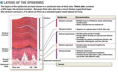
The epidermis is organized into distinct layers, each with specialized functions and cell types.
Stratum | Characteristics |
|---|---|
Stratum basale | Deepest layer; contains stem cells (basal cells), melanocytes, and Merkel cells; forms epidermal ridges. |
Stratum spinosum | 8-10 layers of keratinocytes; contains Langerhans cells; some cells continue dividing. |
Stratum granulosum | 3-5 layers; keratinocytes produce keratin and keratohyalin; cells begin to die. |
Stratum lucidum | Present only in thick skin; clear, glassy layer of densely packed keratin-filled cells. |
Stratum corneum | 15-30 layers of dead, keratinized cells; provides protection and water resistance. |
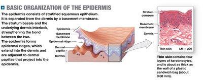
Epidermal Ridges and Dermal Papillae
Epidermal Ridges: Interlock with dermal papillae, increasing surface area for attachment.
Function: Strengthens bond between epidermis and dermis; forms fingerprints.
Dermis
Layers of the Dermis
Papillary Layer: Superficial; areolar tissue; contains capillaries, lymphatics, and sensory neurons.
Reticular Layer: Deep; dense irregular connective tissue; rich in collagen and elastic fibers.
Tension Lines (Cleavage Lines)
Definition: Patterns formed by parallel bundles of collagen and elastic fibers.
Clinical Significance: Incisions parallel to tension lines heal better with less scarring.
Hypodermis (Subcutaneous Layer)
Structure and Function
Composition: Primarily adipose tissue.
Function: Stabilizes skin, stores energy, provides insulation.
Clinical Application: Site for subcutaneous injections.
Epidermal Pigmentation
Types of Pigments
Melanin: Produced by melanocytes; primary determinant of skin color; protects against UV radiation.
Carotene: Orange-yellow pigment from diet; can be converted to vitamin A.
Melanin Distribution
Light-Skinned Individuals: Melanosomes destroyed in superficial layers; less persistent pigmentation.
Dark-Skinned Individuals: Larger, more numerous melanosomes; pigmentation persists in more layers.
Albinism: Normal melanocyte distribution, but no melanin production.
Vitamin D3 Synthesis
Pathway and Importance
UV Radiation: Stimulates production of vitamin D3 (cholecalciferol) in epidermal cells.
Conversion: Liver converts cholecalciferol to calcifediol; kidneys convert calcifediol to calcitriol.
Calcitriol: Essential for calcium and phosphate absorption in the intestine; deficiency leads to rickets.
Accessory Structures
Hair
Structure: Nonliving, cylindrical; produced in hair follicles originating in the dermis.
Layers: Medulla (core), cortex (intermediate), cuticle (surface).
Functions: Protection from UV, sensory reception, prevents entry of foreign particles.
Arrector Pili Muscle: Smooth muscle causing "goose bumps" when contracted.
Exocrine Glands
Sebaceous Glands: Secrete sebum; lubricates hair and skin, inhibits bacteria.
Sweat Glands: Apocrine (thick, odorous sweat; puberty; nutrient for bacteria) and Eccrine (watery sweat; thermoregulation; antimicrobial properties).
Nails
Structure: Dead, keratinized epidermal cells; protect tips of fingers and toes.
Parts: Nail body, lunula, nail root, eponychium (cuticle), hyponychium.
Function: Protection, support during grasping and running.
Summary Table: Integumentary System Components and Functions
Component | Main Function |
|---|---|
Epidermis | Protection, water resistance, sensory reception |
Dermis | Support, blood supply, sensory information |
Hair Follicles | Protection, sensory input |
Exocrine Glands | Excretion, thermoregulation, antimicrobial action |
Nails | Protection, support |
Key Terms and Definitions
Keratin: Tough, fibrous protein produced by keratinocytes.
Melanin: Pigment produced by melanocytes; protects against UV.
Calcitriol: Hormone essential for calcium absorption.
Sebum: Oily secretion from sebaceous glands.
Arrector Pili: Smooth muscle attached to hair follicles.
Important Equations
Vitamin D3 Synthesis Pathway:
Additional info: Academic context was added to clarify the functions, cell types, and clinical significance of the integumentary system, as well as to expand on the structure and function of each layer and accessory structure.
