 Back
BackChapter 5: The Integumentary System - Structured Study Notes
Study Guide - Smart Notes
 Tailored notes based on your materials, expanded with key definitions, examples, and context.
The Integumentary System
Functions of the Skin
The skin is the largest organ of the body and serves multiple essential functions for human health and survival.
Protection: Acts as a barrier against mechanical injury, pathogens, and harmful substances.
Thermoregulation: Regulates internal body temperature through sweat production and blood flow.
Cutaneous Sensation: Contains sensory receptors for touch, pain, temperature, and pressure.
Vitamin D Synthesis: Initiates synthesis of vitamin D when exposed to sunlight, which is necessary for calcium absorption.
Blood Reservoir: Stores blood that can be redirected to other organs as needed.
Excretion and Absorption: Excretes waste products and absorbs certain substances.
Protective Elements of Skin
The skin's protective function is supported by several components:
Keratin: A tough, fibrous protein produced by keratinocytes, providing mechanical strength.
Lipids: Prevent dehydration by reducing water loss.
Glandular Secretions: Sebum and sweat help inhibit microbial growth.
Melanin: Pigment that absorbs UV radiation, protecting deeper tissues.
Thermoregulation
Skin helps maintain a stable internal temperature by controlling heat loss and sweat production.
Vasodilation: Increases blood flow to the skin, promoting heat loss.
Vasoconstriction: Reduces blood flow, conserving heat.
Sweat Glands: Produce sweat, which cools the body as it evaporates.
Vitamin D Synthesis
Exposure to sunlight triggers the conversion of precursor molecules in the skin to vitamin D, which is essential for calcium absorption and bone health.
Importance: Without vitamin D, dietary calcium cannot be absorbed efficiently.
Structure of the Skin
Layers of the Skin
The skin consists of two main layers and an underlying subcutaneous layer:
Epidermis: The superficial, avascular layer composed of epithelial cells.
Dermis: The deeper, vascular layer made of connective tissue.
Hypodermis: Not technically part of the skin; consists of adipose tissue and anchors skin to underlying structures.
Cell Types in the Epidermis
The epidermis contains four primary cell types:
Keratinocytes: Make up 90% of epidermal cells; produce keratin.
Melanocytes: Produce melanin pigment; comprise 8% of cells.
Langerhans Cells: Immune cells that help defend against pathogens.
Merkel Cells: Sensory cells involved in touch perception.
Epidermal Layers (Deep to Superficial)
The epidermis is organized into five distinct layers:
Stratum Basale: Deepest layer; site of mitosis; contains all four cell types.
Stratum Spinosum: Contains keratinocytes and Langerhans cells; melanocyte production occurs here.
Stratum Granulosum: Contains lamellar granules; apoptosis (programmed cell death) occurs.
Stratum Lucidum: Present only in thick skin (e.g., palms, soles).
Stratum Corneum: Outermost layer; consists of keratinized cells that are continuously shed.
Dermis
The dermis is the thick, supportive layer beneath the epidermis, containing blood vessels, nerves, and connective tissue.
Papillary Layer: Made of areolar connective tissue; contains dermal papillae (finger-like projections).
Reticular Layer: Thickest layer; composed of dense, irregular connective tissue.
Hypodermis
The hypodermis, or subcutaneous layer, is not part of the skin but provides insulation and energy storage.
Composition: Mainly adipose tissue.
Function: Anchors skin to underlying structures.
Skin Color
Factors Affecting Skin Color
Skin color is determined by three main pigments:
Hemoglobin: Red pigment in blood; oxygenated blood gives skin a pinkish hue.
Carotene: Yellow-orange pigment from diet; accumulates in stratum corneum.
Melanin: Brown-black pigment produced by melanocytes; protects against UV radiation.
Skin Color Disorders
Albinism: Genetic condition where melanin is not produced.
Vitiligo: Loss of melanocytes in patches, resulting in depigmented areas.
Accessory Structures of the Skin
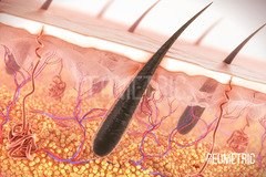
Hair
Hair serves protective and sensory functions and is composed of several anatomical parts.
Function: Protection from UV rays, minor trauma, and light touch sensation.
Anatomy: Shaft, root, hair bulb, hair papilla, matrix, arrector pili muscle.
Shaft: Composed of medulla, cortex, and cuticle.
Root: Includes internal and external root sheaths, and dermal root sheath.
Hair Follicle: Surrounds the root and anchors the hair.
Alopecia: Rapid hair loss in defined areas; caused by genetics, endocrine disorders, chemotherapy, or stress.
Skin Exocrine Glands
The skin contains several types of exocrine glands, each with distinct functions:
Sebaceous Glands: Simple acinar, holocrine glands; secrete sebum into hair follicles.
Sudoriferous (Sweat) Glands: Include merocrine (eccrine) and apocrine glands.
Merocrine (Eccrine) Glands: Simple tubular; found in palms, soles, forehead; regulate temperature and remove waste.
Apocrine Glands: Simple tubular; found in armpits and genital regions; viscous secretions activated by stress or sexual arousal.
Ceruminous (Wax) Glands: Simple tubular, merocrine; found in external auditory canal; produce cerumen (earwax).
Sebum
Function: Prevents hair from drying out, keeps skin soft, reduces water loss.
Cerumen
Function: Forms a sticky barrier to prevent entry of foreign bodies into the ear.
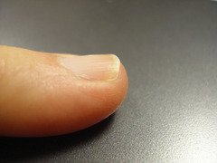
Nails
Nails are protective coverings composed of tightly packed, keratinized cells.
Nail Body: Visible portion; includes free edge, nail body, lunula, eponychium (cuticle), and nail root (not visible).
Lunula: White, crescent-shaped area; thickened stratum basale.
Nail Root: Located beneath the skin.
Eponychium: Cuticle; protects the nail matrix.
Skin Injuries and Disorders
Burns
Burns are classified by depth and severity:
First Degree Burn: Affects only the epidermis; causes redness and pain (e.g., sunburn).
Second Degree Burn: Damages dermis and epidermis; causes blistering.
Third Degree Burn: Destroys all skin layers; requires skin grafting; results in charring.
Rule of Nines
The Rule of Nines is used to estimate the extent and severity of burns by dividing the body into regions, each representing approximately 9% of total body surface area.
Purpose: Guides treatment and fluid replacement decisions.
Summary Table: Skin Layers and Their Features
Layer | Main Features | Cell Types |
|---|---|---|
Stratum Basale | Deepest, mitosis, single cell layer | Keratinocytes, Melanocytes, Langerhans, Merkel |
Stratum Spinosum | Several cell layers, spiny appearance | Keratinocytes, Langerhans |
Stratum Granulosum | Granules, apoptosis | Keratinocytes |
Stratum Lucidum | Clear, only in thick skin | Keratinocytes |
Stratum Corneum | Outermost, keratinized, shed | Keratinocytes |
Dermis | Vascular, connective tissue | Fibroblasts, immune cells |
Hypodermis | Adipose, anchors skin | Adipocytes |
