 Back
BackChapter 5: The Integumentary System – Study Notes
Study Guide - Smart Notes
 Tailored notes based on your materials, expanded with key definitions, examples, and context.
The Integumentary System
Overview and Introduction
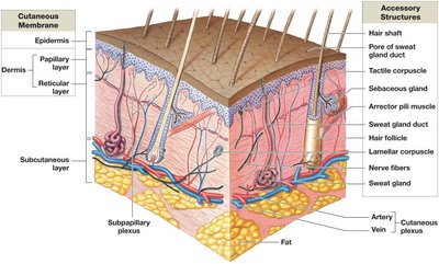
The integumentary system is the largest organ system of the human body, serving as the primary barrier between internal tissues and the external environment. It consists of the cutaneous membrane (skin) and various accessory structures. The system plays a critical role in protection, sensation, thermoregulation, and metabolic functions.
Cutaneous membrane: Composed of the epidermis (superficial epithelium) and dermis (deep connective tissue).
Accessory structures: Include hair, hair follicles, exocrine glands, and nails.
Subcutaneous layer (hypodermis): Lies deep to the dermis, separating the skin from underlying organs.
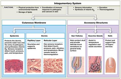
Functions of the Integumentary System
Protection: Shields underlying tissues from mechanical damage, pathogens, and dehydration.
Excretion: Removes salts, water, and organic wastes via exocrine glands.
Thermoregulation: Maintains body temperature through insulation and evaporative cooling.
Melanin production: Protects against ultraviolet (UV) radiation.
Keratin production: Provides toughness and water resistance.
Vitamin D3 synthesis: Essential for calcium metabolism.
Sensory reception: Detects touch, pressure, temperature, and pain.
Structure of the Skin
Cutaneous Membrane
Epidermis: Keratinized, stratified squamous epithelium; avascular; relies on diffusion from dermal capillaries.
Dermis: Deep connective tissue layer containing blood vessels, nerves, and accessory structures.
Subcutaneous layer (hypodermis): Loose connective and adipose tissue; not technically part of the skin but stabilizes it.
Accessory Structures
Hair and hair follicles
Exocrine glands (sebaceous and sweat glands)
Nails
The Epidermis
General Features
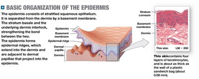
The epidermis is composed primarily of keratinocytes, which produce the protein keratin. It is organized into distinct layers (strata) and is responsible for the protective barrier function of the skin.
Keratinocytes: Main cell type; produce keratin for strength and water resistance.
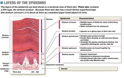
Types of skin: Thin skin (four layers) covers most of the body; thick skin (five layers) is found on palms and soles.

Layers of the Epidermis (from deep to superficial)
Stratum basale (germinativum): Deepest layer; contains stem cells (basal cells) that divide to produce new keratinocytes; attached to basement membrane by hemidesmosomes; contains melanocytes and tactile (Merkel) cells.
Stratum spinosum: 8–10 layers of keratinocytes bound by desmosomes; contains dendritic (Langerhans) cells for immune defense.
Stratum granulosum: 3–5 layers; keratinocytes stop dividing, produce keratin and keratohyalin; cells become flatter and die.
Stratum lucidum: Present only in thick skin; clear, densely packed, keratin-filled cells.
Stratum corneum: 15–30 layers of dead, keratinized cells; forms the exposed surface; cells are shed after about two weeks.
Epidermal Ridges and Surface Patterns
The stratum basale forms epidermal ridges that interlock with dermal papillae, increasing the surface area for attachment and forming the basis for fingerprints.
Specialized Cells of the Epidermis
Merkel (tactile) cells: Sensory receptors for touch, found in the stratum basale of hairless skin.
Melanocytes: Produce melanin pigment; processes extend into superficial layers.
Dendritic (Langerhans) cells: Immune cells in the stratum spinosum.
The Dermis
Structure and Layers
The dermis is a connective tissue layer deep to the epidermis, anchoring accessory structures and providing strength and elasticity to the skin.
Papillary layer: Superficial; composed of areolar tissue; contains capillaries, lymphatics, and sensory neurons; forms dermal papillae.
Reticular layer: Deep; dense irregular connective tissue rich in collagen and elastic fibers; provides strength and flexibility.
Tension lines (cleavage lines): Patterns of collagen and elastic fibers; incisions parallel to these lines heal better with less scarring.
Clinical Note: Dermatitis
Inflammation of the dermis, especially the papillary layer; can be caused by infection, chemicals, or irritation.
The Hypodermis (Subcutaneous Layer)
The hypodermis is primarily composed of adipose tissue and serves as an energy reserve, insulator, and shock absorber. It is the site for subcutaneous injections.
Epidermal Pigmentation
Melanin and Carotene
Melanin: Produced by melanocytes in the stratum basale; protects against UV radiation; stored in melanosomes and transferred to keratinocytes.
Carotene: Orange-yellow pigment from diet; accumulates in epidermal cells and can be converted to vitamin A.
Skin color differences are due to the amount and persistence of melanin, not the number of melanocytes. Albinism results from the inability to produce melanin.
Vitamin D3 Synthesis
UV radiation stimulates the production of vitamin D3 (cholecalciferol) in the skin, which is converted by the liver and kidneys to calcitriol, a hormone essential for calcium and phosphate absorption in the intestine. Deficiency can lead to rickets in children.
Accessory Structures of the Skin
Hair
Produced in hair follicles; protects against UV radiation, insulates, and provides sensory input.
Structure: Medulla (core), cortex (middle), cuticle (surface).
Arrector pili muscle causes hair to stand (goosebumps).
Exocrine Glands
Sebaceous glands: Secrete sebum (oil) into hair follicles; lubricates and protects skin and hair.
Sebaceous follicles: Discharge sebum directly onto skin surface.
Apocrine sweat glands: Secrete thick, odorous sweat into hair follicles; found in armpits, nipples, pubic region; active at puberty.
Eccrine (merocrine) sweat glands: Secrete watery sweat directly onto skin; important for thermoregulation and antimicrobial defense.
Nails
Composed of dead, keratinized cells; protect finger and toe tips; aid in manipulation and defense.
Key structures: Nail body, lunula, nail root, eponychium (cuticle), hyponychium.
