 Back
BackChapter 5: The Skeletal System – Structure and Function of the Upper Limb Bones
Study Guide - Smart Notes
 Tailored notes based on your materials, expanded with key definitions, examples, and context.
The Skeletal System
Introduction to the Skeletal System
The skeletal system provides the structural framework for the human body, supporting movement, protecting internal organs, storing minerals, and facilitating blood cell formation. Bones are classified by their tissue type and shape, and their structure is adapted to their function.
Support: Bones form the rigid structure that supports the body and maintains its shape.
Protection: Bones protect vital organs, such as the skull protecting the brain and the rib cage protecting the heart and lungs.
Movement: Bones act as levers for muscles, enabling movement at joints.
Storage: Bones store minerals, primarily calcium and phosphorus, which are released into the bloodstream as needed.
Blood Cell Formation: Hematopoiesis occurs in the red bone marrow, producing red and white blood cells.
Classification of Bones
Bones are classified based on their tissue type and shape:
Osseous Tissue: Compact bone (dense and strong) and spongy bone (porous and lightweight).
Shape: Long bones (e.g., femur), short bones (e.g., carpals), flat bones (e.g., skull), and irregular bones (e.g., vertebrae).
Bones of the Upper Limb
Structure of the Hand
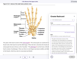
The human hand consists of three main groups of bones: phalanges, metacarpals, and carpals. These bones allow for a wide range of movements and dexterity.
Phalanges: The fingers are composed of three phalanges each (proximal, middle, distal), except for the thumb, which has two (proximal and distal).
Metacarpals: Five metacarpal bones form the palm of the hand, numbered I to V from the thumb to the little finger.
Carpals: Eight carpal bones form the wrist, arranged in two rows. The proximal row includes the scaphoid, lunate, triquetrum, and pisiform; the distal row includes the trapezium, trapezoid, capitate, and hamate.
Example: The arrangement of carpal bones allows for the flexibility and strength required for gripping and manipulating objects.
Labeling the Bones of the Hand
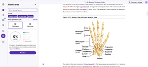
Understanding the anatomy of the hand is essential for identifying bone injuries and for clinical assessments. The labeled diagram below shows the anterior view of the right hand, highlighting the phalanges, metacarpals, and carpals.
Key Terms and Definitions
Phalanges: The bones of the fingers and toes.
Metacarpals: The five bones forming the palm of the hand.
Carpals: The eight bones forming the wrist.
Radius and Ulna: The two bones of the forearm that articulate with the carpal bones.
Clinical Relevance
Knowledge of the hand's bone structure is crucial for diagnosing fractures, dislocations, and other injuries. For example, the scaphoid bone is commonly fractured due to falls on an outstretched hand.
Summary Table: Bones of the Hand
Bone Group | Number of Bones | Location | Example Bone |
|---|---|---|---|
Phalanges | 14 | Fingers | Distal phalanx |
Metacarpals | 5 | Palm | Metacarpal I (thumb) |
Carpals | 8 | Wrist | Scaphoid |
Additional info:
These notes expand upon the brief points in the original material, providing definitions, examples, and clinical context for the bones of the upper limb. The images included are directly relevant, showing labeled diagrams of the hand bones for anatomical study.
