 Back
BackChapter 5: The Skeletal System – Structure and Function of the Upper Limb Bones
Study Guide - Smart Notes
 Tailored notes based on your materials, expanded with key definitions, examples, and context.
The Skeletal System
Introduction to the Skeletal System
The skeletal system is a vital organ system that provides structural support, protection for internal organs, facilitates movement, stores minerals, and is the site of blood cell formation. Bones are classified by their tissue type and shape, and their structure is closely related to their function.
Support: Bones form the framework that supports the body and cradles soft organs.
Protection: Bones protect vital organs such as the brain, heart, and lungs.
Movement: Bones act as levers for muscles, enabling movement.
Storage: Bones store minerals, primarily calcium and phosphorus.
Blood Cell Formation: Hematopoiesis occurs in the red marrow of certain bones.
Classification of Bones
Bones can be classified based on their shape and the type of osseous tissue present.
Compact Bone: Dense and solid, forms the outer layer of bones.
Spongy Bone: Porous and lightweight, found inside bones.
Long Bones: Longer than they are wide (e.g., femur, humerus).
Short Bones: Cube-shaped (e.g., carpals, tarsals).
Flat Bones: Thin and flattened (e.g., skull, ribs).
Irregular Bones: Complex shapes (e.g., vertebrae, pelvis).
Bones of the Upper Limb
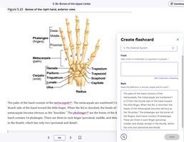
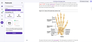
Structure of the Hand (Figure 5.23)
The human hand consists of three main groups of bones: phalanges, metacarpals, and carpals. These bones allow for a wide range of movement and dexterity.
Phalanges: The bones of the fingers; each finger has three phalanges (proximal, middle, distal), except the thumb, which has two.
Metacarpals: The five bones forming the palm of the hand.
Carpals: Eight bones forming the wrist, arranged in two rows.
Carpal Bones: The carpal bones are arranged in two rows:
Proximal row: Scaphoid, Lunate, Triquetrum, Pisiform
Distal row: Trapezium, Trapezoid, Capitate, Hamate
The radius and ulna are the forearm bones that articulate with the carpal bones to form the wrist joint.
Bone Markings
Bone markings are anatomical features on bones that serve as sites for muscle attachment, passage of nerves and blood vessels, and articulation with other bones. Examples include:
Foramen: A hole for passage of blood vessels or nerves.
Process: A projection for muscle attachment.
Condyle: A rounded articular surface.
Joints of the Upper Limb
Joints, also known as articulations, are connections between bones that allow for movement and provide stability. The hand contains several important joints:
Carpometacarpal Joint: Between the carpal and metacarpal bones.
Metacarpophalangeal Joint: Between the metacarpal and phalangeal bones.
Interphalangeal Joint: Between the phalanges.
Clinical Relevance
Understanding the anatomy of the hand is essential for diagnosing fractures, dislocations, and other injuries. The arrangement of bones and joints allows for complex movements such as gripping, pinching, and fine motor tasks.
Example: A fracture of the scaphoid bone is common in falls onto an outstretched hand and may require careful management due to its poor blood supply.
Summary Table: Bones of the Hand
Bone Group | Number of Bones | Location | Function |
|---|---|---|---|
Phalanges | 14 | Fingers | Movement, dexterity |
Metacarpals | 5 | Palm | Support, structure |
Carpals | 8 | Wrist | Flexibility, articulation |
Additional info:
Flashcards and matching study tools are useful for memorizing bone names, locations, and functions. Practice labeling diagrams and identifying bone features to reinforce learning.
