 Back
Chapter 6: Bone and Skeletal Tissue – Structured Study Notes
Study Guide - Smart Notes
 Tailored notes based on your materials, expanded with key definitions, examples, and context.
Bone and Skeletal Tissue
Overview and Importance
The skeletal system provides the structural framework for the human body, supports movement, and protects vital organs. Understanding bone anatomy and physiology is essential for diagnosing and treating bone diseases such as osteoporosis.
Skeletal Cartilages
Basic Structure and Types
Skeletal cartilage is the first tissue of the skeleton, providing flexibility and resilience. It is gradually replaced by bone during development but remains in areas where flexibility is needed.
No blood vessels or nerves: Cartilage relies on diffusion for nutrient supply.
Surrounded by perichondrium: A dense connective tissue layer.
Contains chondrocytes in lacunae: Cells embedded in a gel-like extracellular matrix.
Types of Cartilage
Hyaline cartilage: Provides support and forms joints; most common type.
Elastic cartilage: Highly flexible; found in the ear and epiglottis.
Fibrocartilage: Strong and acts as a shock absorber; found in intervertebral discs and knee menisci.
Cartilage Locations
Cartilage is found in joints, rib attachments, ear, nose, and respiratory structures.
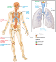
Cartilage Growth and Calcification
Appositional growth: Cartilage grows outward from the perichondrium.
Interstitial growth: Cartilage grows from within, as chondrocytes divide.
Calcification: Occurs during growth and aging, making cartilage stiff and prone to damage. This is not the same as bone formation.
Functions of Bones
Major Functions
Support: Provides structural support for the body and soft organs.
Protection: Shields the brain, spinal cord, and vital organs.
Anchorage: Acts as levers for muscle action.
Mineral and growth factor storage: Reservoir for calcium, phosphorus, and growth factors.
Blood cell formation: Hematopoiesis occurs in red marrow cavities.
Triglyceride storage: Fat stored in bone cavities serves as an energy source.
Hormone production: Osteocalcin regulates insulin secretion, glucose levels, and metabolism.
Skeletal Organization
Axial vs. Appendicular Skeleton
Axial skeleton: Skull, vertebral column, rib cage.
Appendicular skeleton: Limbs and girdles.
Classification of Bones
Bone Shapes
Long bones: Longer than wide (e.g., femur).
Short bones: Cube-shaped (e.g., carpals).
Sesamoid bones: Embedded within tendons (e.g., patella).
Flat bones: Thin and protective (e.g., skull, ribs).
Irregular bones: Complex shapes (e.g., vertebrae).
Bone Structure Overview
Types of Bone Tissue
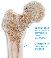
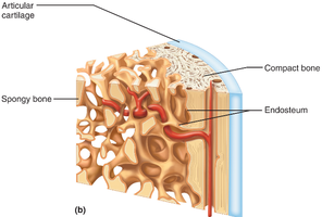
Compact bone: Dense, outer layer providing strength.
Spongy bone: Lattice structure (trabeculae) containing marrow, lightweight but strong.
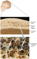
Structure of Short, Irregular, and Flat Bones
Spongy bone (diploë) inside, covered by compact bone.
Periosteum (outer layer) and endosteum (inner lining).
No central cavity.
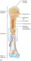
Long Bone Anatomy
Diaphysis: Shaft of the bone.
Epiphyses: Ends of the bone.
Medullary cavity: Contains marrow.
Articular cartilage: Covers joint surfaces.
Epiphyseal line/plate: Site of growth.
Membranes, Blood Supply, Marrow, and Markings
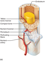
Periosteum: Covers outer bone surface; fibrous and osteogenic layers; contains blood vessels and nerves; anchors tendons and ligaments.
Endosteum: Lines internal bone surfaces; covers trabeculae; contains bone-forming cells.
Blood supply: Nutrient arteries enter through foramina; nerves travel with vessels.
Bone Marrow
Red marrow: Site of blood cell formation; more common in children.
Yellow marrow: Fat storage; predominates in adults, especially in long bones.
Bone Markings
Attachment sites for muscles and ligaments.
Joint surfaces.
Openings for vessels and nerves.
Types of Bone Markings
Projections: Attachment points.
Surfaces: Joint formation.
Openings: Passage for vessels and nerves.
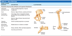
Bone Markings: Attachments
Name of Bone Marking | Description | Illustrations |
|---|---|---|
Crest | Narrow ridge of bone; usually prominent | Hip bone |
Tubercle | Small rounded projection or process | Femur |
Spine | Sharp, slender, often pointed projection | Vertebra |
Process | Any bony prominence | Spinous process |
Trochanter | Very large, blunt, irregularly shaped process | Femur |
Line | Narrow ridge of bone; less prominent than a crest | Intertrochanteric line |
Tuberosity | Large rounded projection; may be roughened | Ischial tuberosity |
Bone Markings: Surfaces and Openings
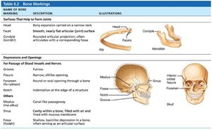
Name of Bone Marking | Description | Illustrations |
|---|---|---|
Head | Bony expansion carried on a narrow neck | Femur head |
Facet | Smooth, nearly flat articular surface | Vertebra facet |
Condyle | Rounded articular projection | Mandible condyle |
Foramen | Round or oval opening through a bone | Skull foramen |
Meatus | Canal-like passageway | Skull meatus |
Fossa | Shallow depression in a bone | Skull fossa |
Microscopic Anatomy of Bone
Bone Cells
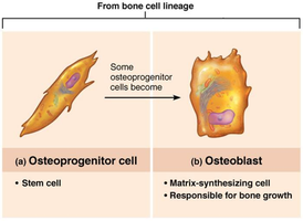
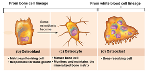
Osteoprogenitor cells: Stem cells found in periosteum and endosteum; differentiate into osteoblasts.
Osteoblasts: Build bone by secreting osteoid and promoting mineralization; actively mitotic.
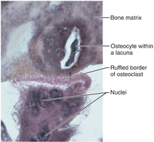
Osteocytes: Mature bone cells; maintain bone matrix and monitor stress.
Bone-lining cells: Cover bone surfaces; help maintain matrix.
Osteoclasts: Large, multinucleated cells responsible for bone resorption.
Compact Bone Organization
Osteon (Haversian system): Structural unit of compact bone; consists of concentric lamellae around a central canal.
Lamellae: Collagen fibers in alternating directions resist twisting.
Canals and canaliculi: Central (Haversian) canals contain blood vessels and nerves; perforating (Volkmann’s) canals connect central canals; canaliculi are tiny channels for nutrient and signal exchange.
Lamellae types: Interstitial (between osteons) and circumferential (around bone).
Spongy Bone Microscopy
Trabeculae arranged along stress lines; no osteons; lightweight but strong.
Bone Composition
Organic and Inorganic Components
Organic components: Osteoid (collagen and proteins) and bone cells; provide tensile strength and flexibility.
Inorganic components: Hydroxyapatites (mineral salts, mainly calcium and phosphate); provide hardness and resistance to compression.
Bone Development (Ossification)
Types of Ossification
Endochondral ossification: Replaces hyaline cartilage; most bones form this way; begins in the diaphysis.
Intramembranous ossification: Bone forms directly from mesenchymal tissue; flat bones (skull, clavicle).
Endochondral Ossification Steps
Bone collar forms around cartilage model.
Cartilage calcifies and cavities form.
Spongy bone forms.
Epiphyses ossify; cartilage remains at joints and growth plates.
Intramembranous Ossification Steps
Osteoblasts form from mesenchymal cells.
Osteoid is secreted and calcifies.
Trabeculae form, creating spongy bone.
Bone matures.
Postnatal Bone Growth
Growth in Length and Width
Length: Occurs at the epiphyseal plate; stops when plates close (~18 years in females, ~21 in males).
Width: Appositional growth; osteoblasts add bone outside, osteoclasts remove inside.
Growth Plate Zones
Proliferation: Cells divide.
Hypertrophic: Cells enlarge.
Calcification: Cells die.
Ossification: Bone forms.
Hormonal Regulation of Bone Growth
Growth hormone: Stimulates bone growth.
Thyroid hormone: Ensures proper proportions.
Sex hormones: Cause growth spurt and plate closure.
Bone Remodeling
Process and Regulation
Continuous process; about 5–10% of bone mass recycled each year.
Spongy bone replaced every 3–4 years; compact bone every 10 years.
Bone deposit (osteoblasts) and resorption (osteoclasts) maintain strength and calcium balance.
Hormonal Controls
Parathyroid hormone (PTH): Increases blood calcium by stimulating osteoclasts.
Calcitonin: Decreases blood calcium by inhibiting osteoclasts.
Vitamin D: Essential for calcium absorption.
Mechanical Stress (Wolff’s Law)
Bones adapt to mechanical stress; more stress leads to stronger bone.
Osteocytes detect stress and signal osteoblasts to build more bone.
Bone Repair
Fracture Classification
Position: Displaced vs. nondisplaced.
Completeness: Complete vs. incomplete.
Skin: Open vs. closed.
Fracture Treatment and Repair
Hematoma formation: Blood clot forms at fracture site.
Fibrocartilaginous callus formation: Soft callus forms, stabilizing the fracture.
Bony callus formation: Hard callus replaces soft callus.
Bone remodeling: Bone is reshaped to original form.
Bone Disorders
Osteomalacia and Rickets
Caused by vitamin D or calcium deficiency.
Poor mineralization leads to soft, weak bones.
Osteoporosis
Bone resorption exceeds deposition; bone mass decreases.
Increased fracture risk.
Risk factors: aging, postmenopausal status, low activity, poor diet, smoking, alcohol, genetics, certain medications, hyperthyroidism, diabetes mellitus.
Prevention: weight-bearing exercise, calcium and vitamin D, medications to reduce resorption.
Paget’s Disease
Abnormal remodeling; rapid, disorganized bone formation.
Results in weak bone structure.
Additional info: These notes expand upon the original content with academic context, definitions, and examples to ensure completeness and clarity for exam preparation.
