 Back
BackChapter 6: Bone Structure and Function – Study Notes
Study Guide - Smart Notes
 Tailored notes based on your materials, expanded with key definitions, examples, and context.
Bone Structure and Function
Overview of the Skeletal System
The skeletal system is composed of bones, cartilages, ligaments, and other connective tissues that stabilize and connect different elements. It provides movement, structural support, protection, storage of minerals and lipids, and blood cell production.
Movement and Locomotion: Bones act as levers for muscles.
Structural Support and Attachment: Provides framework for the body and attachment points for muscles.
Protection: Protects vital organs (e.g., thoracic cage, vertebrae, skull).
Storage: Stores calcium (most abundant ion in the body) and lipids (in yellow bone marrow).
Blood Cell Production: Occurs in red bone marrow.
Bone Structure
Long Bones vs. Flat Bones
Bones are classified by shape and internal structure. Long bones and flat bones have distinct anatomical features.
Long Bones: Consist of a diaphysis (shaft), metaphysis (narrow zone), and epiphysis (ends). The diaphysis contains compact bone and a medullary cavity with yellow marrow. The epiphysis contains spongy bone with red marrow.
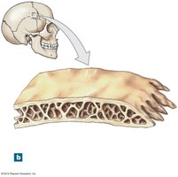
Flat Bones: Have a core of spongy bone (diploë) with red marrow, sandwiched between layers of compact bone.
Osseous Tissue and Bone Matrix
Composition and Properties
Bone tissue (osseous tissue) is a specialized connective tissue with a dense, mineralized matrix and collagen fibers. The matrix is composed of:
Hydroxyapatite crystals: Calcium phosphate (Ca3(PO4)2) and calcium hydroxide (Ca(OH)2).
Calcium carbonate (CaCO3): Adds compressive strength but is brittle.
Collagen fibers: Provide tensile strength and flexibility.
The combination of minerals and collagen makes bone tough and lightweight, capable of being remodeled.
Cell Types in Bone
Osteoprogenitor Cells
Osteoprogenitor cells are mesenchymal stem cells that differentiate into osteoblasts. They are found in the inner cellular layer of the periosteum and endosteum and assist in fracture repair.
Osteoblasts
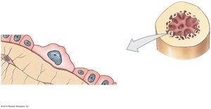
Osteoblasts are immature bone cells responsible for producing new bone matrix (osteogenesis). They secrete osteoid, which later becomes calcified. Once surrounded by bone, osteoblasts become osteocytes.
Osteocytes
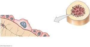
Osteocytes are mature bone cells that maintain the bone matrix. They reside in lacunae and connect via canaliculi. Osteocytes do not divide but can become osteoblasts during bone repair.
Osteoclasts
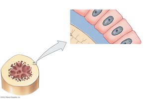
Osteoclasts are large, multinucleated cells derived from monocyte/macrophage lineage. They secrete acids and proteases to dissolve bone matrix (osteolysis), releasing stored minerals and weakening bone. Osteoclasts and osteoblasts work in balance to remodel bone.
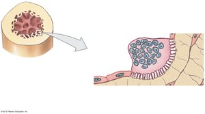
Structure of Compact and Spongy Bone
Compact Bone
Compact bone is organized into osteons, the basic structural units. Osteocytes are arranged in concentric lamellae around a central canal containing blood vessels. Perforating canals run perpendicular to the central canal, connecting osteons.
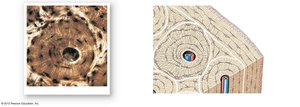
Spongy Bone
Spongy bone consists of a network of trabeculae, with spaces filled by red bone marrow. It lacks osteons, and nutrients reach osteocytes by diffusion through canaliculi. In some bones, yellow marrow stores fat.
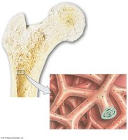
The Periosteum and Endosteum
The Periosteum
The periosteum is a dense irregular connective tissue covering the exterior of all compact bones except at joint capsules. It consists of an outer fibrous layer and an inner cellular layer. Perforating fibers anchor the periosteum to bone, ligaments, and tendons.
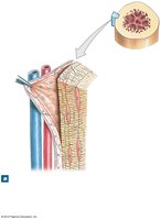
The Endosteum
The endosteum lines the medullary cavity, central canals, and covers trabeculae of spongy bone. It contains osteoblasts, osteoprogenitor cells, and osteoclasts, and is active in bone growth and repair.
Bone Development and Ossification
Ossification Processes
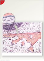
Bone formation (osteogenesis or ossification) begins at 6 weeks of embryonic development and continues until about age 25. Two main types of ossification are:
Endochondral Ossification: Most bones form from hyaline cartilage templates. The process involves the formation of a primary ossification center in the diaphysis, followed by secondary centers in the epiphyses. Growth in length continues at the epiphyseal cartilage until puberty.
Intramembranous (Dermal) Ossification: Produces flat bones like the mandible, clavicle, and cranial bones. Mesenchymal cells differentiate into osteoblasts, forming bone directly without a cartilage stage.
Blood and Nerve Supply in Bones
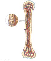
Bones are highly vascularized, containing arteries, veins, lymphatic vessels, and sensory nerves. The nutrient artery and vein enter the diaphysis through the nutrient foramen. Metaphyseal and periosteal vessels supply other regions. After epiphyseal closure, the vascular network becomes interconnected.
Bone Repair and Remodeling
Bone Fracture Repair
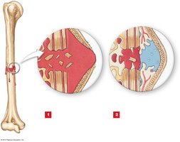
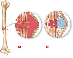
Bone repair involves several stages:
Hematoma Formation: Bleeding forms a clot at the fracture site.
Callus Formation: An external callus (cartilage and bone) and internal callus (bone) stabilize the fracture. Fibroblasts and osteoprogenitor cells contribute to new tissue formation.
Bone Formation: Cartilage in the callus is replaced by bone.
Remodeling: Spongy bone is replaced by compact bone, restoring the bone's original shape.
Bone Remodeling and Exercise
Bone is continuously remodeled by the coordinated activity of osteocytes, osteoblasts, and osteoclasts. About 20% of the skeleton is remodeled each year. Mechanical stress and exercise stimulate bone deposition, making bones stronger, while inactivity leads to bone loss.
Hormonal Regulation of Bone; Parathyroid Hormone (PTH)
Hormonal and Nutritional Effects
Bone acts as a reservoir for calcium, phosphate, and magnesium. Hormones regulate bone growth and mineral balance:
Estrogens and Androgens: Stimulate osteoblasts and bone growth.
Thyroxine and Growth Hormone: Stimulate metabolism and bone growth.
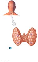
Parathyroid Hormone (PTH): Increases plasma Ca2+ by stimulating osteoclasts, reducing renal excretion, and increasing intestinal absorption (via calcitriol).
Calcitonin: Decreases plasma Ca2+, but has a minor role in humans.
PTH and Calcium Homeostasis
PTH is secreted when plasma Ca2+ falls below 2.1 mM. It acts on bone, kidneys, and intestines to restore normal calcium levels through a negative feedback loop.
Bone Aging and Disease
Osteopenia and Osteoporosis
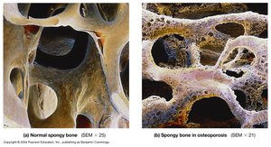
With age, bones become thinner and weaker due to decreased osteoblast activity. Osteopenia begins between ages 30 and 40, progressing to osteoporosis if bone loss compromises function. Women are more affected due to decreased estrogen after menopause.
Factor | Effect on Bone |
|---|---|
Estrogen/Androgen | Stimulate osteoblasts, maintain bone mass |
PTH | Increases blood Ca2+, stimulates osteoclasts |
Calcitonin | Decreases blood Ca2+, inhibits osteoclasts (minor role) |
Exercise | Increases bone strength and mass |
Aging | Decreases osteoblast activity, increases risk of osteoporosis |
Example: Osteoporosis is a disease characterized by decreased bone mass and increased fracture risk, especially in postmenopausal women due to reduced estrogen levels.
